


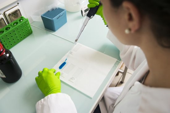
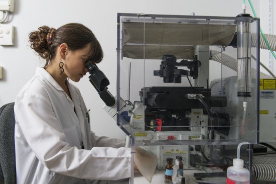
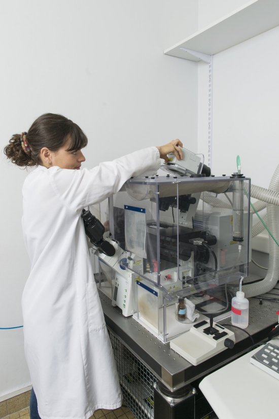

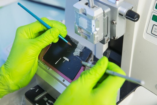
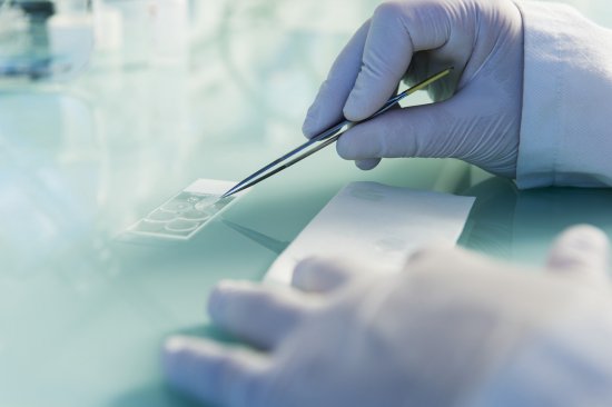
Photo
20160102_0028
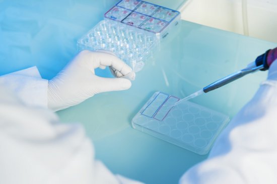
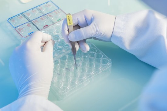
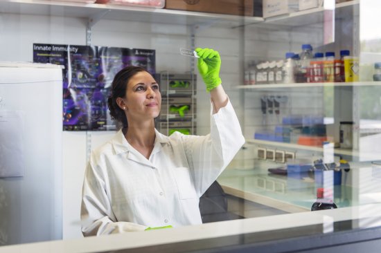
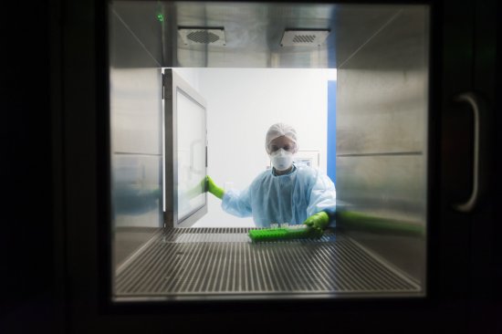
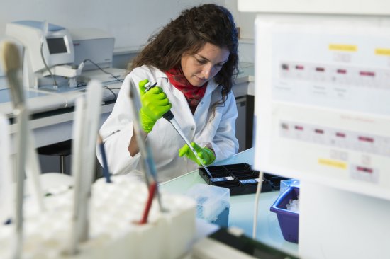
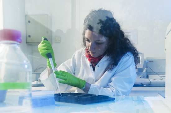
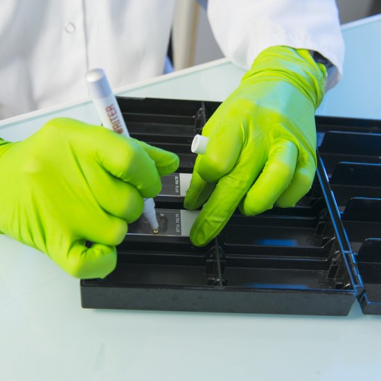
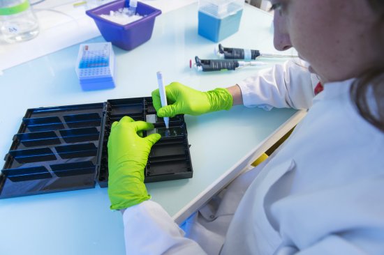
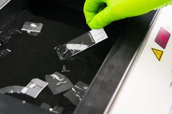
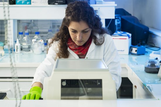
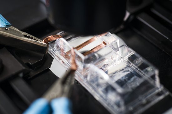
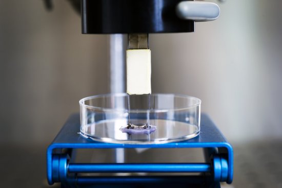
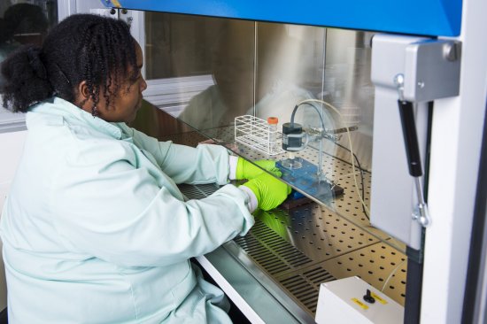
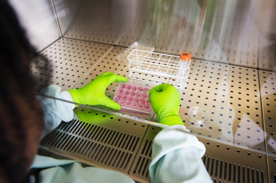
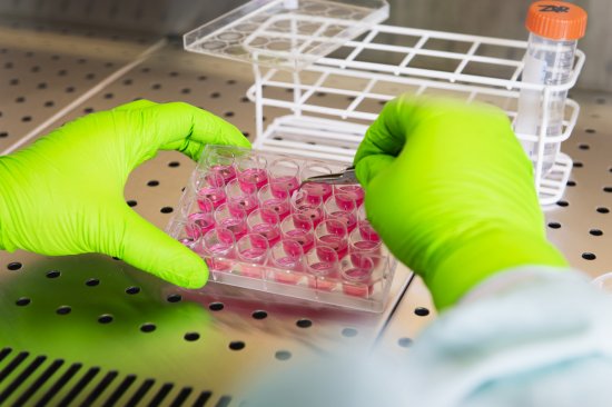
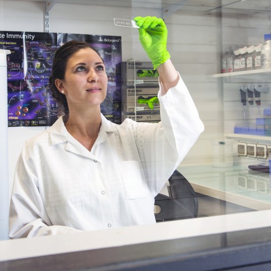
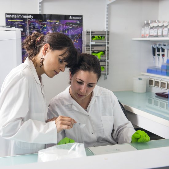
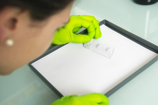
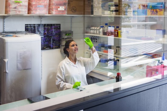
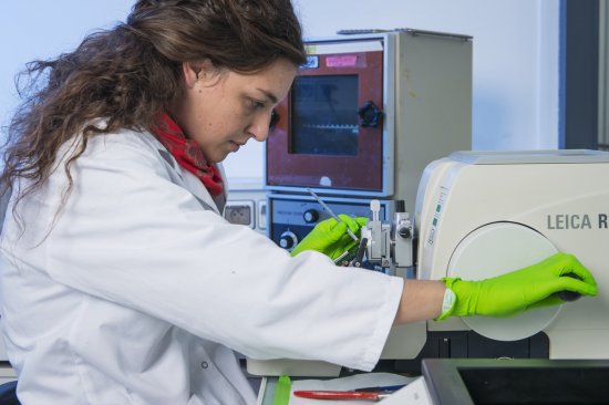
Fixing cells infected by HIV and stained for immunofluorescence on a glass slide
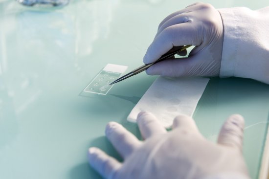
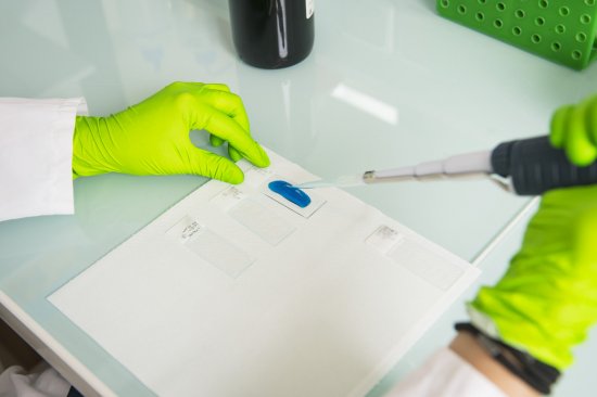
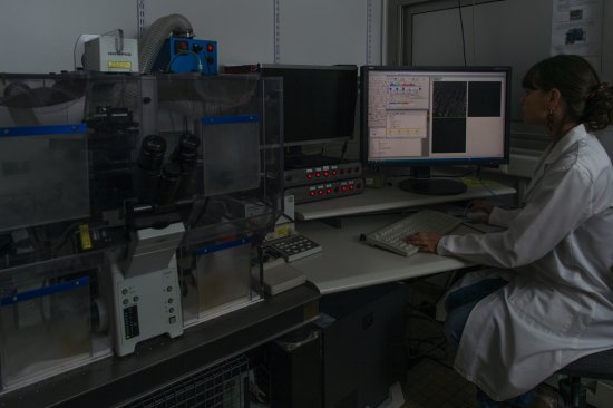
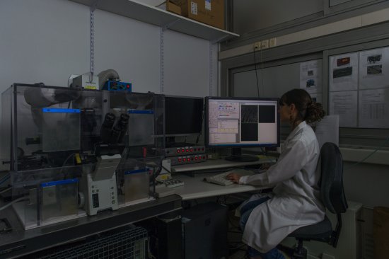
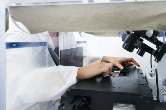
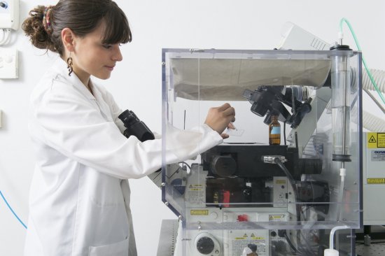
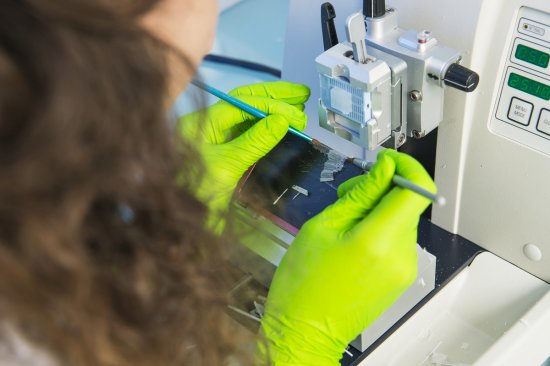
Photo
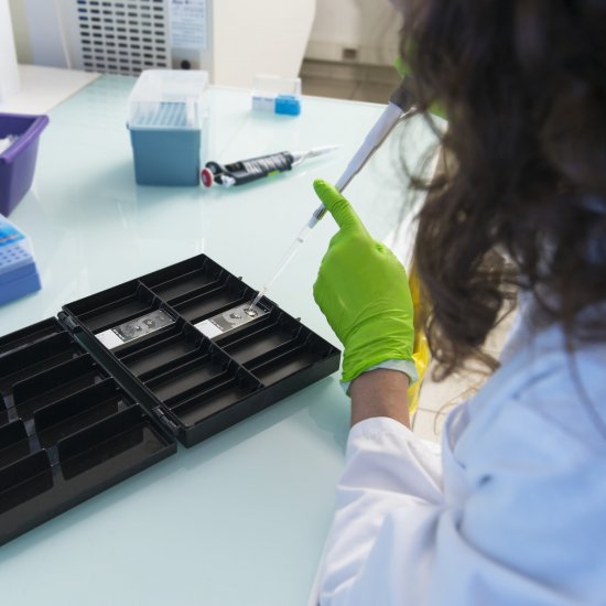
20160102_0027
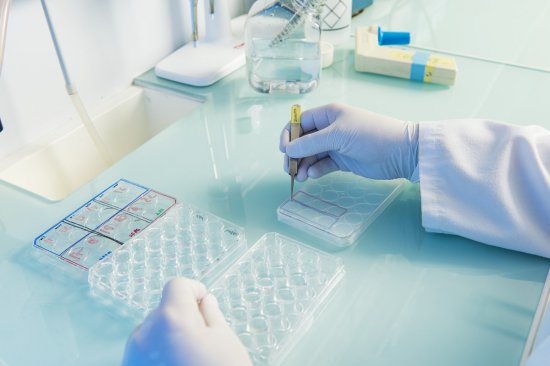
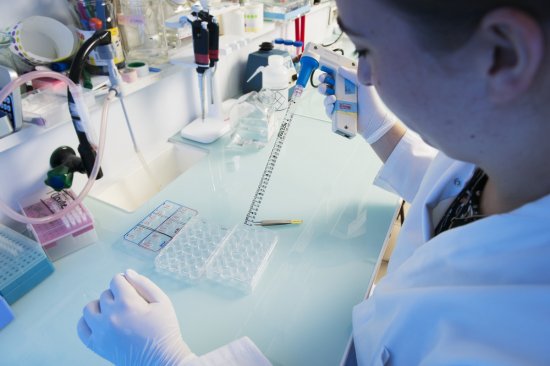
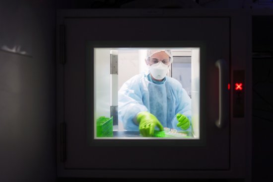
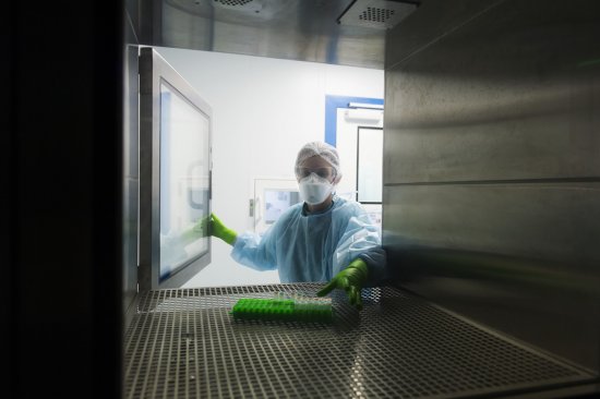
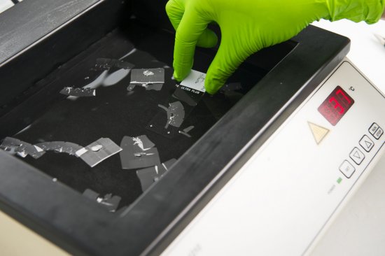
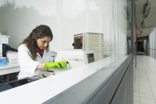
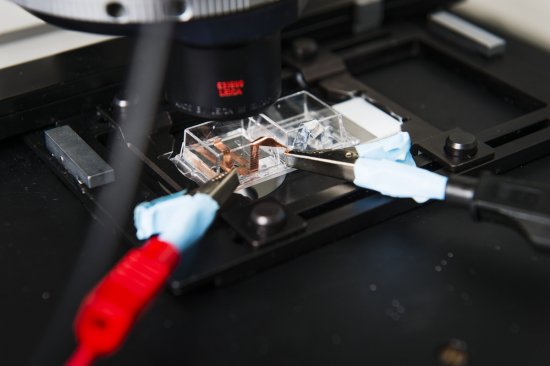
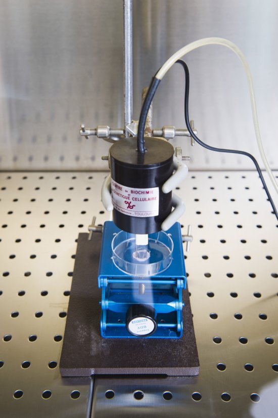
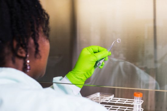
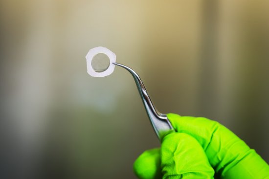
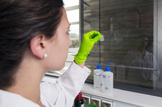
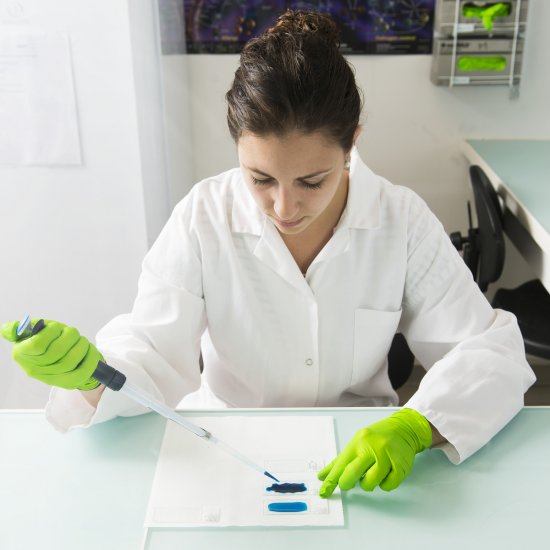
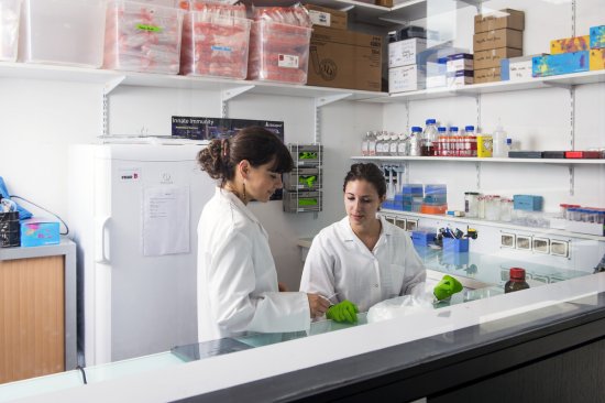
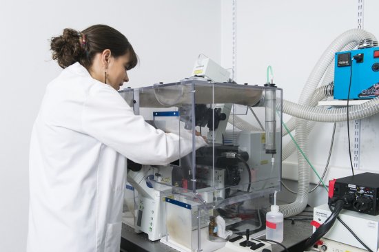
Fixing cells infected by HIV and stained for immunofluorescence on a glass slide