Production year
2016

© Cyril FRESILLON/IPBS/CNRS Images
20160102_0067
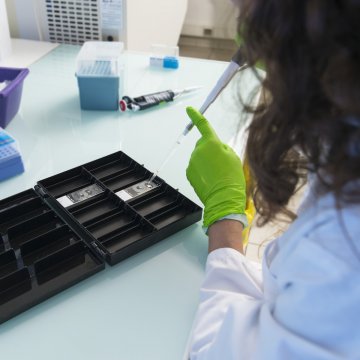
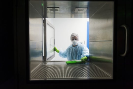
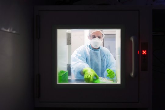
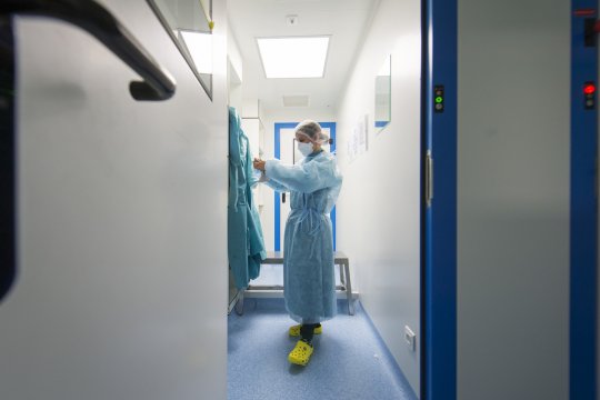
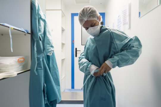
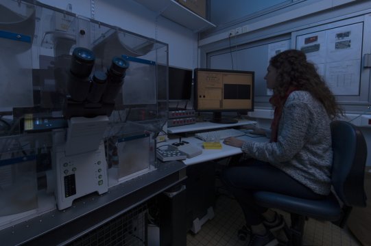
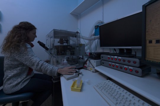
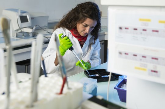
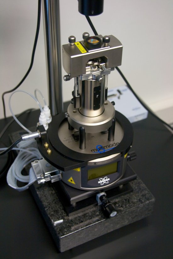
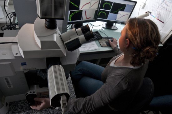
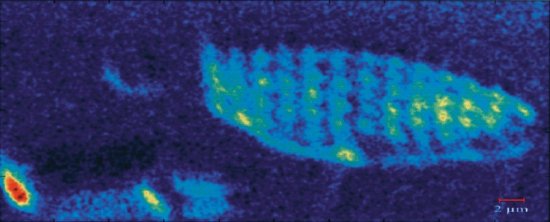
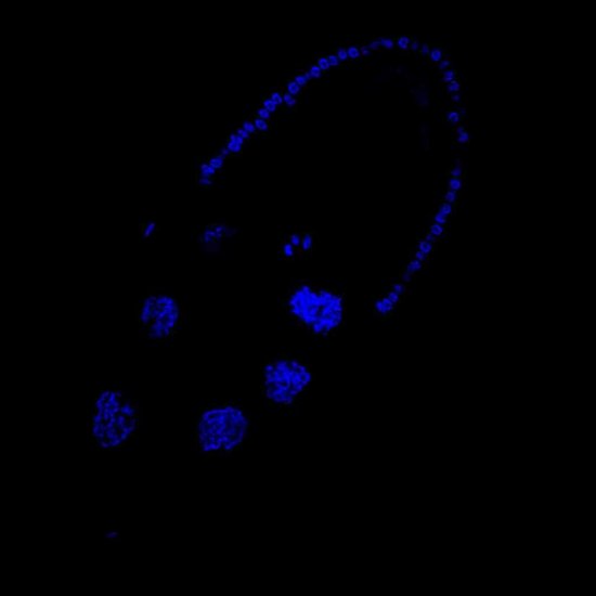
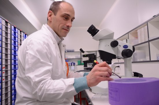
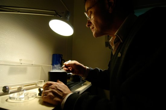
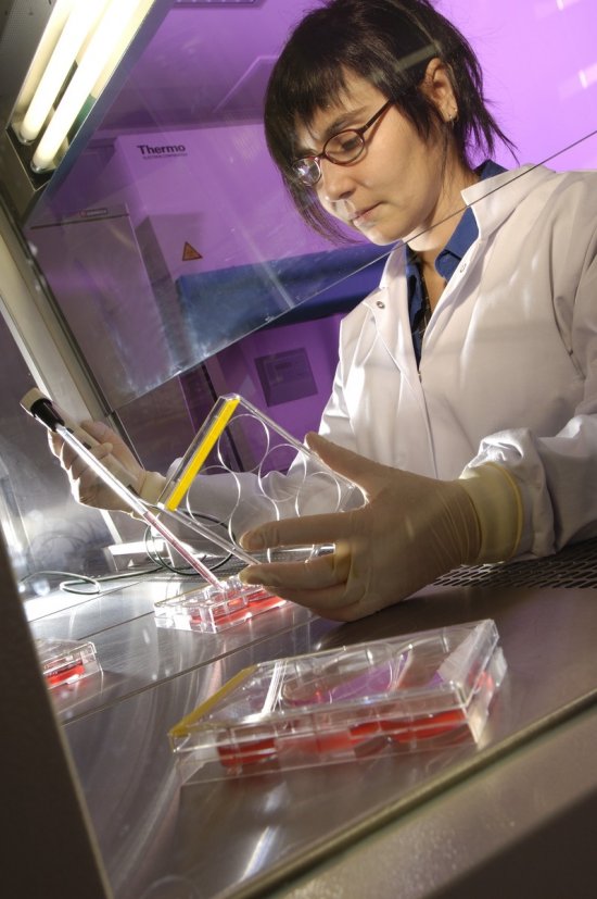
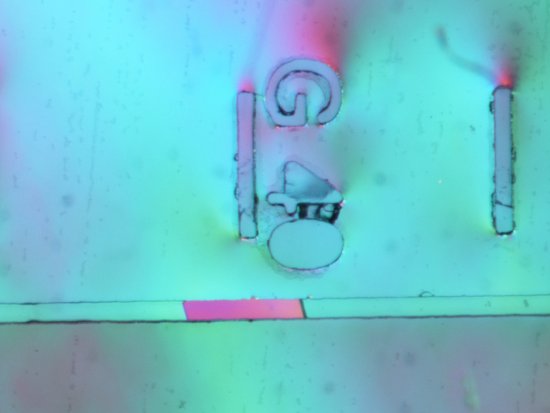
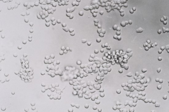
Application of a pulsed electric field to a slice of human dermal tissue. This tissue is a dermal substitute engineered from cells taken during a skin biopsy. The tissue was placed between electrodes connected to a generator (in yellow). The generator transmitted electrical impulses with known parameters to induce a transitory permeabilisation of the cell membranes of the tissue and enable a GFP-expressing plasmid (Green Fluorescent Protein, a protein that emits fluorescent green light) to penetrate the cells. After 24 or 48 hours, the tissue was examined by multiphoton microscope in order to locate and quantify the amount of GFP-expressing cells. This experiment enabled us to obtain in vitro rates of transfection comparable to those obtained in vivo. It thus proved possible to create a substitute for human skin in vitro, i.e. a dermal model that can be used in the laboratory. This will allow us to limit the number of in vivo tests on animal models.
The use of media visible on the CNRS Images Platform can be granted on request. Any reproduction or representation is forbidden without prior authorization from CNRS Images (except for resources under Creative Commons license).
No modification of an image may be made without the prior consent of CNRS Images.
No use of an image for advertising purposes or distribution to a third party may be made without the prior agreement of CNRS Images.
For more information, please consult our general conditions
2016
Our work is guided by the way scientists question the world around them and we translate their research into images to help people to understand the world better and to awaken their curiosity and wonderment.