Production year
2016

© Cyril FRESILLON/IPBS/CNRS Images
20160102_0023
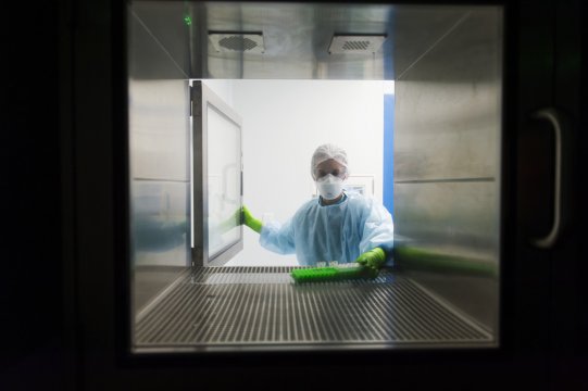
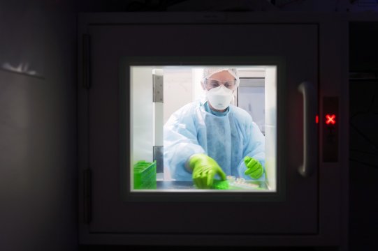
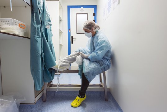
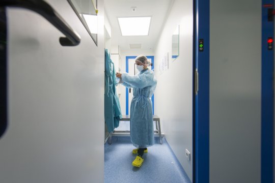
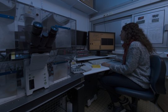
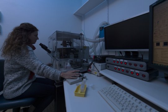
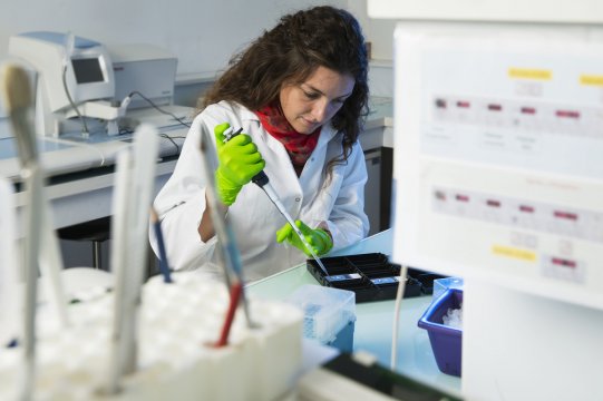
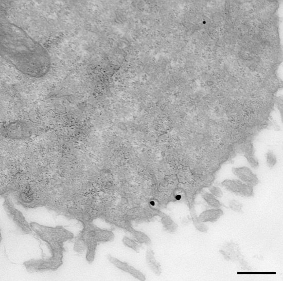
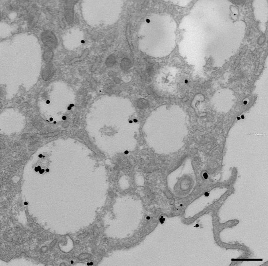
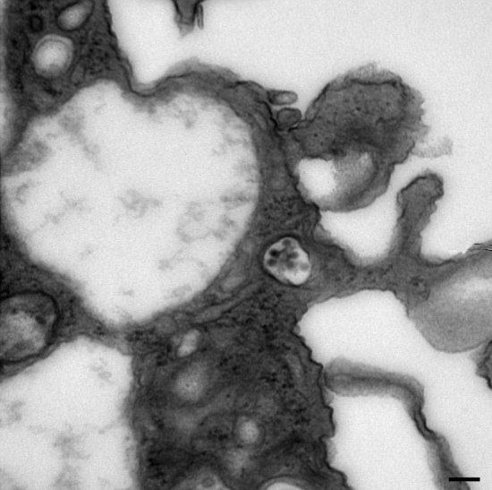
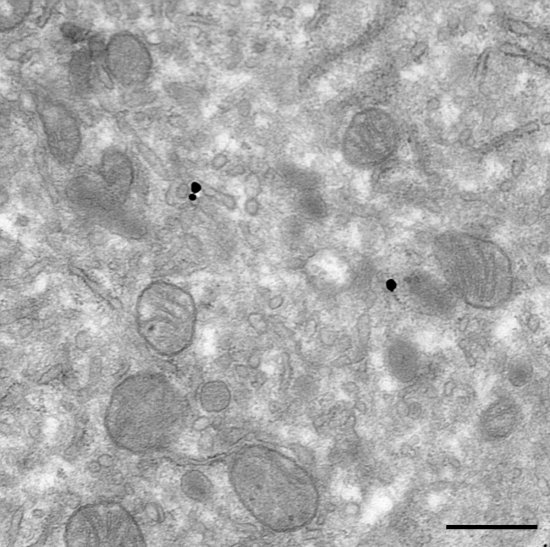
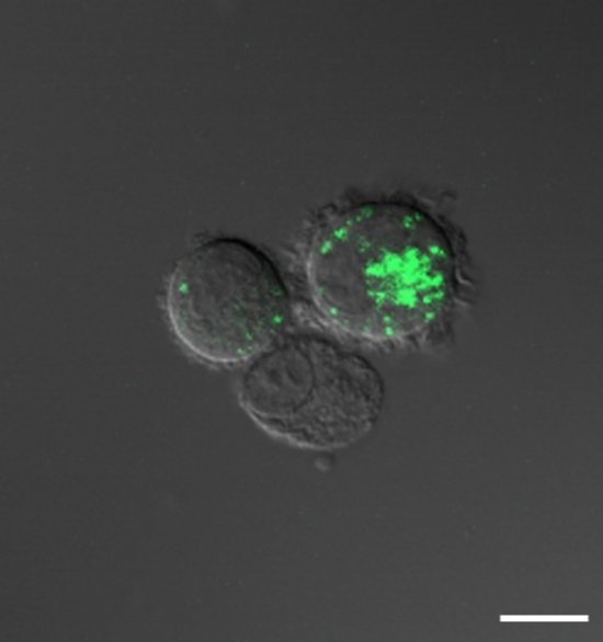
Immunofluorescence staining of cells infected by HIV, previously fixed on a glass slide. Human macrophages, immune cells, were extracted from the blood of healthy donors. These were then put into culture and co-infected with HIV and Mycobacterium tuberculosis, the agent responsible for tuberculosis. The infected cells were stained with visible fluorescent antibodies which specifically recognize HIV. Immunofluorescence is used to track the development of the HIV infection and to understand the mechanisms that explain the aggravation of the infection induced by tuberculosis.
The use of media visible on the CNRS Images Platform can be granted on request. Any reproduction or representation is forbidden without prior authorization from CNRS Images (except for resources under Creative Commons license).
No modification of an image may be made without the prior consent of CNRS Images.
No use of an image for advertising purposes or distribution to a third party may be made without the prior agreement of CNRS Images.
For more information, please consult our general conditions
2016
Our work is guided by the way scientists question the world around them and we translate their research into images to help people to understand the world better and to awaken their curiosity and wonderment.