Production year
2016

© Cyril FRESILLON/IPBS/CNRS Images
20160102_0074
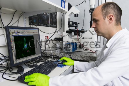
Analysis of fluorescence images of the extracellular matrix of a bacterial biofilm of S. epidermidis, positioned under a fluorescence microscope (in the background). Copper electrodes were connected to a generator by means of two alligator clips. The biofilms were then subjected to pulsed electric fields in order to disorganise the bacteria. The aim was to understand how the extracellular matrix is organised and how the pulsed electric fields can disorganise it. The long term goal is to use pulsed electric fields to combat nosocomial diseases resistant to antibiotics.
The use of media visible on the CNRS Images Platform can be granted on request. Any reproduction or representation is forbidden without prior authorization from CNRS Images (except for resources under Creative Commons license).
No modification of an image may be made without the prior consent of CNRS Images.
No use of an image for advertising purposes or distribution to a third party may be made without the prior agreement of CNRS Images.
For more information, please consult our general conditions
2016
Our work is guided by the way scientists question the world around them and we translate their research into images to help people to understand the world better and to awaken their curiosity and wonderment.