Production year
2016

© Cyril FRESILLON/IPBS/CNRS Images
20160102_0045
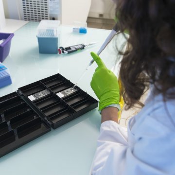
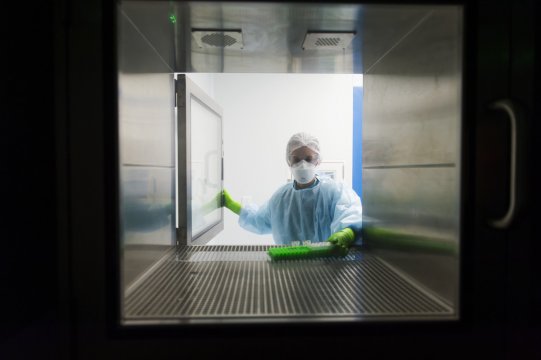
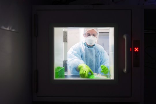
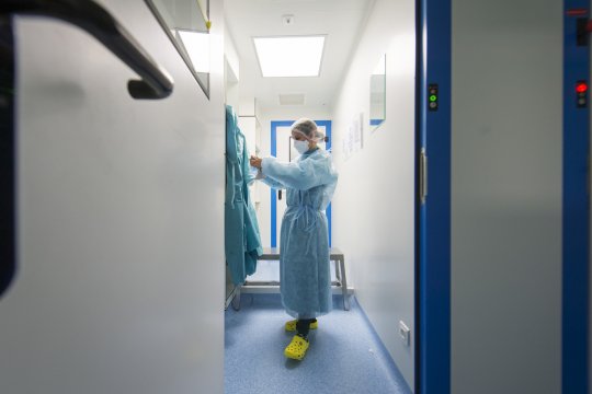
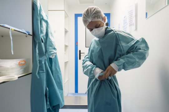
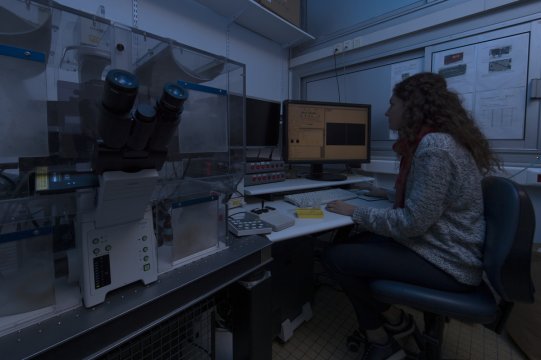
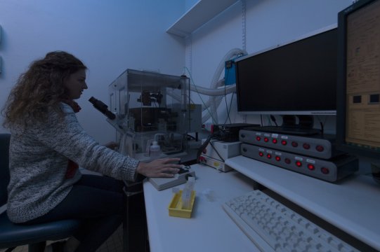
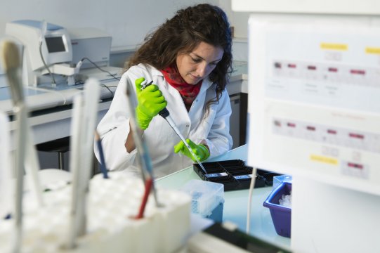
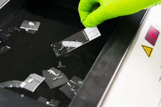
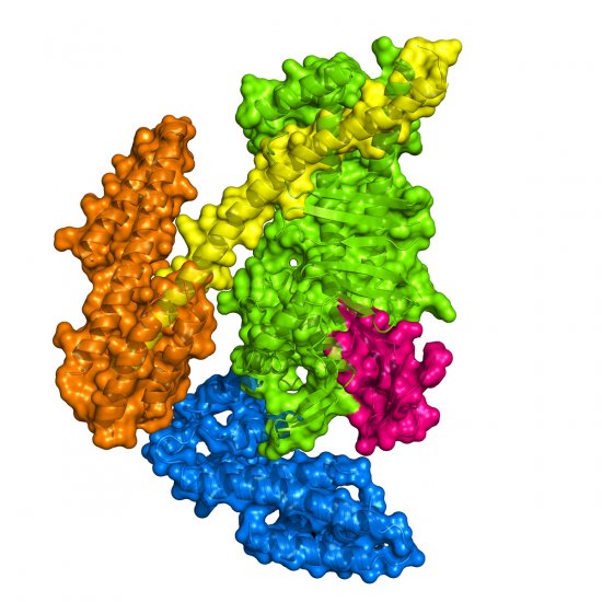
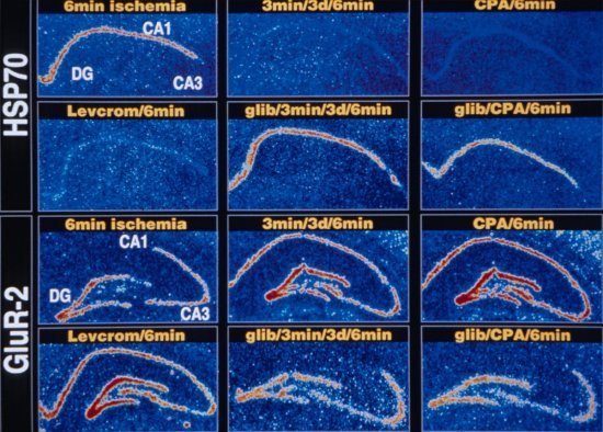
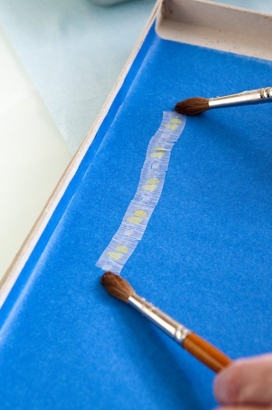
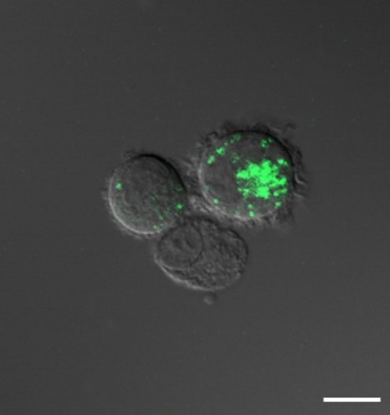
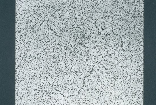
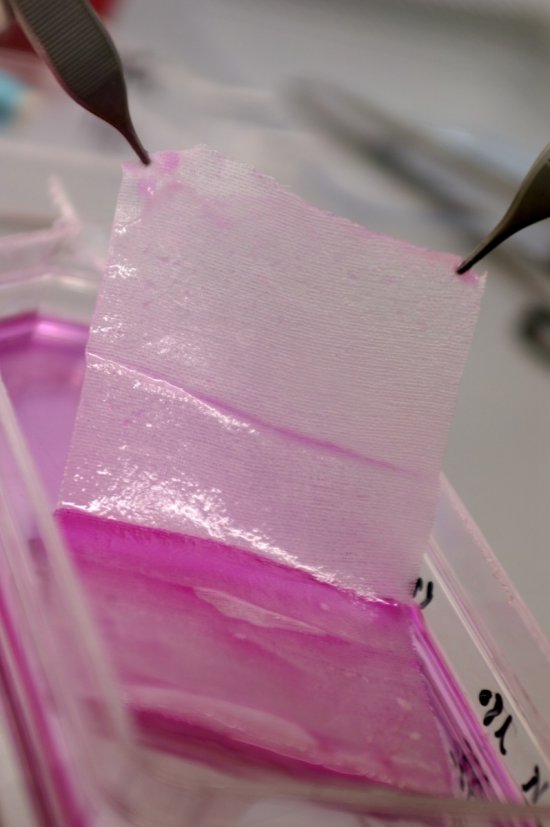
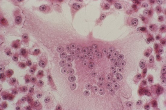
Sections of murine fibrosarcoma induced subcutaneously in mice placed on glass slides were circled using an ImmunoMarker. The fibrosarcoma models spontaneously generated HEV blood vessels (high endothelium veinules) 8 days after induction in the mouse. Researchers were seeking to detect the presence of specific RNA for these blood vessels on the glass slides. They therefore deposited a mixture of antibodies on the sections in order to co-mark the RNA of the vessels. The ImmunoMarker formed a water-repellent barrier around the tumour in order to preserve the antibodies after they have been positioned.
The use of media visible on the CNRS Images Platform can be granted on request. Any reproduction or representation is forbidden without prior authorization from CNRS Images (except for resources under Creative Commons license).
No modification of an image may be made without the prior consent of CNRS Images.
No use of an image for advertising purposes or distribution to a third party may be made without the prior agreement of CNRS Images.
For more information, please consult our general conditions
2016
Our work is guided by the way scientists question the world around them and we translate their research into images to help people to understand the world better and to awaken their curiosity and wonderment.