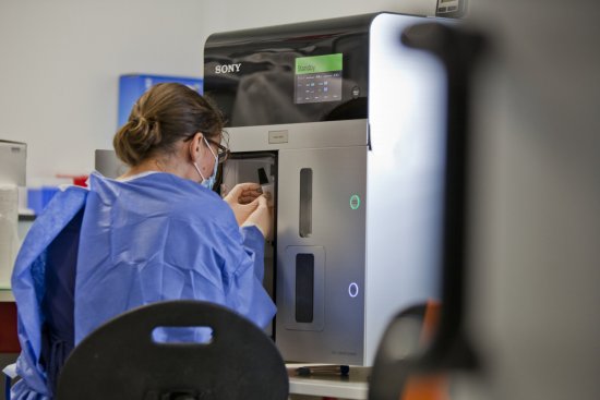
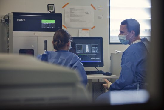
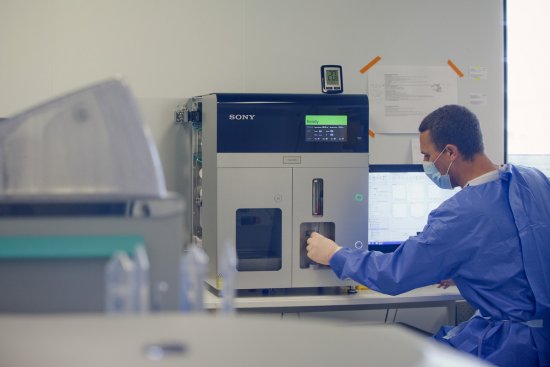
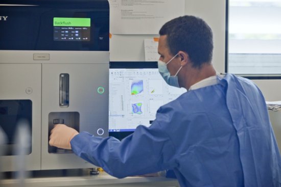
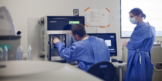
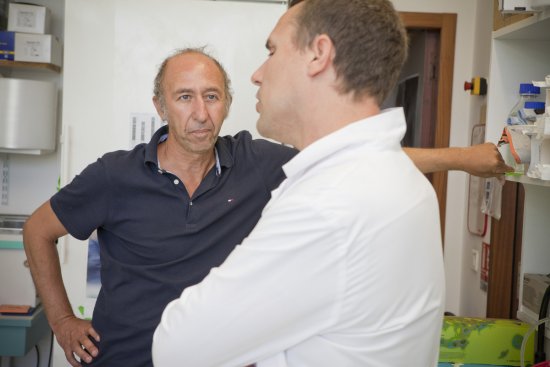
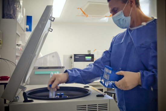
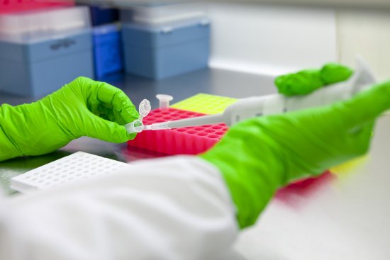
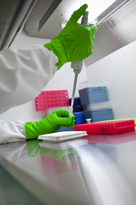
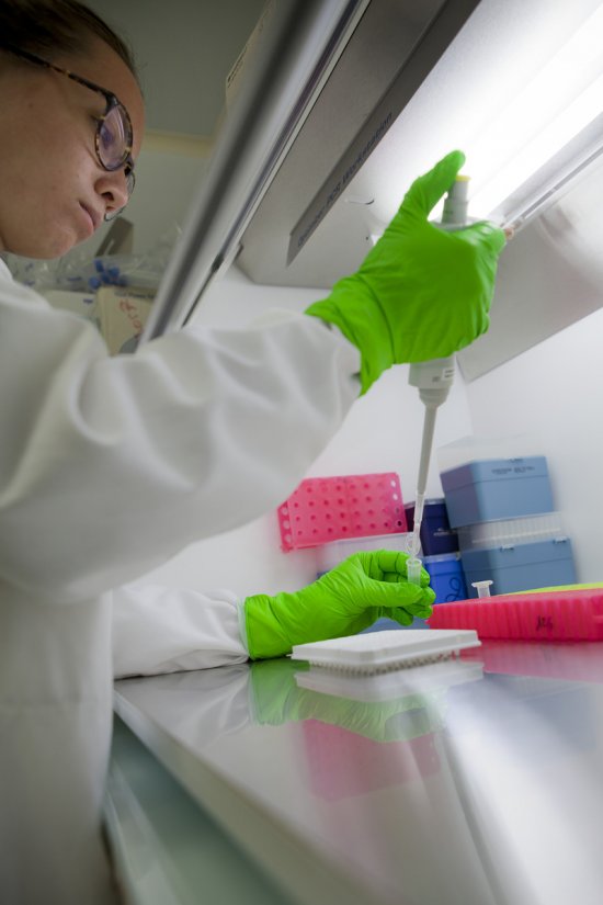
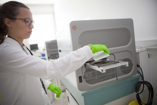
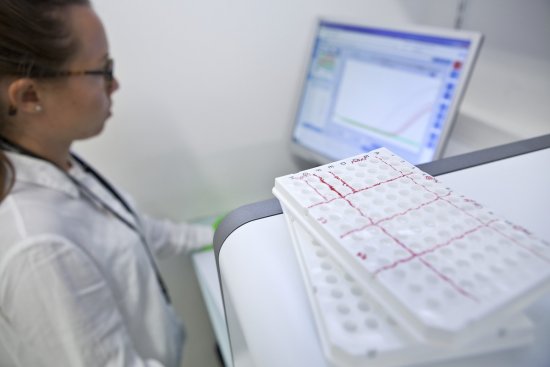
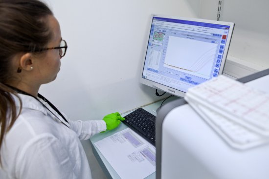
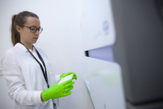
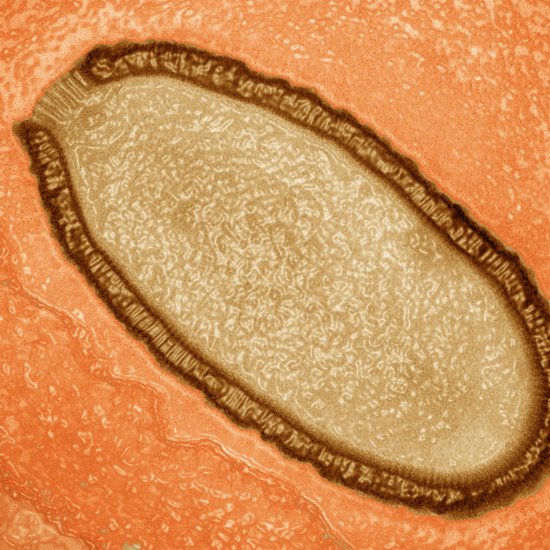
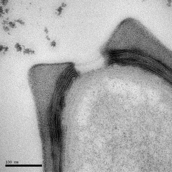
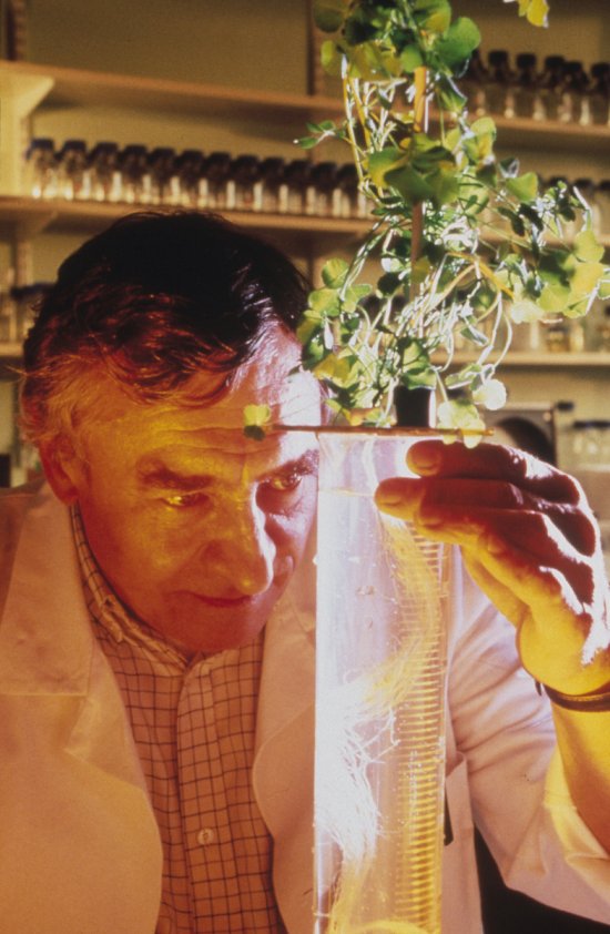
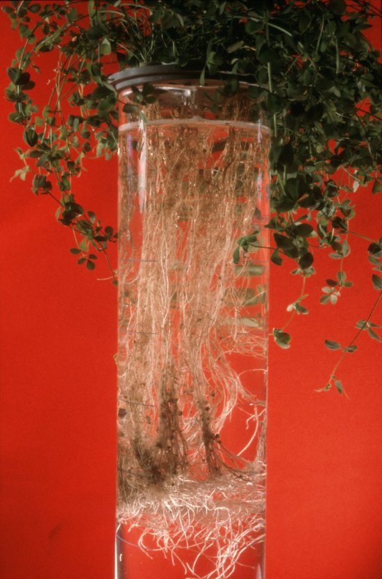
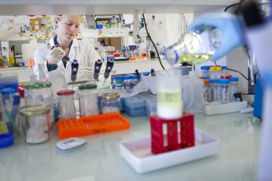
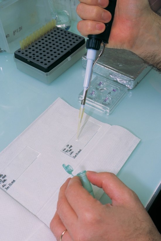
Photo report

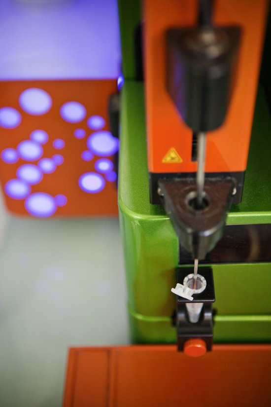
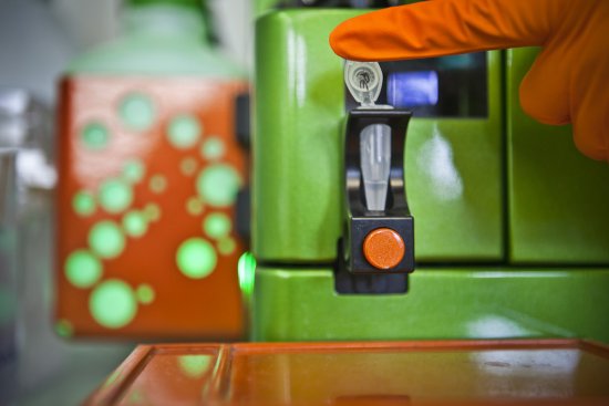
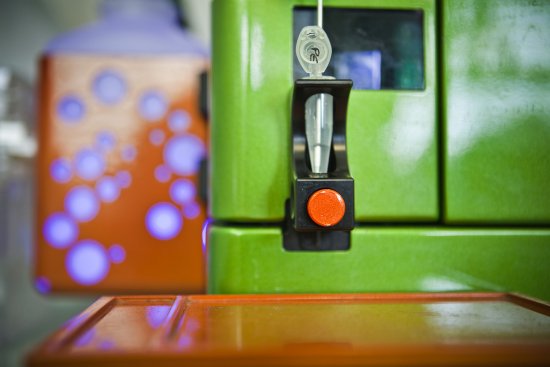
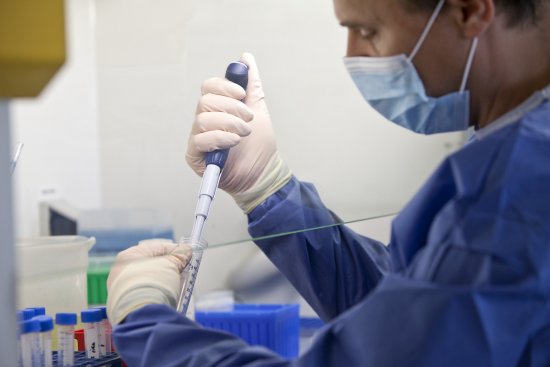
Institut de génétique humaine (IGH)
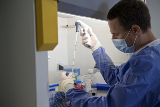
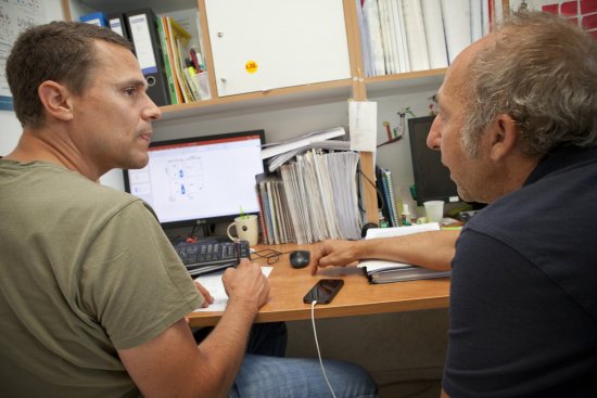
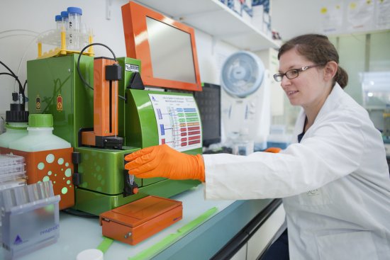
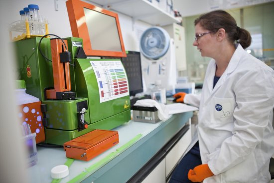
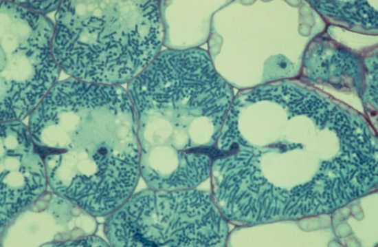
HIV report

20170072_0001
26 media
20170072_0001
Open media modal

Handling a blood sample from a patient infected with HIV-1 in a BSC (biosafety cabinet) in the P3 laboratory at the Institute of Human Genetics (IGH). All the peripheral blood mononuclear cells (PBMC) are collected in order to mark them with a specific antibody. The PBMC ring (formed element of the blood: mainly leucocytes) has been separated from the red blood cells and the plasma. The PBMC contain both healthy cells (the vast majority) and infected cells (very rare). It is in this laboratory…

Photo
20170072_0001
Manipulation d’un prélèvement sanguin de patient infecté par le VIH-1 dans le labo P3 de l'IGH
20170072_0032
Open media modal

Inserting a sample into a FACS (fluorescence-activated cell sorting) machine used to characterise the infected cells (lymphocytes) of patients with the marker CD32+. This machine makes it possible to view the cells and analyse their specific surface markers by using fluorochrome-conjugated antibodies directed against the markers of interest. The aim is to analyse and characterise these CD32+ cells in detail.

Photo
20170072_0032
Insertion d'un échantillon dans une machine de FACS (Fluorescent Activated Cell Sorting)
20170072_0033
Open media modal

FACS (fluorescence-activated cell sorting) machine used to characterise the infected cells (lymphocytes) of patients with the marker CD32+. This machine makes it possible to view the cells and analyse their specific surface markers by using fluorochrome-conjugated antibodies directed against the markers of interest. The aim is to analyse and characterise these CD32+ cells in detail.

Photo
20170072_0033
Machine de FACS utilisée pour caractériser des cellules (lymphocytes) infectées
20170072_0034
Open media modal

Sample in a FACS (fluorescence-activated cell sorting) machine used to characterise the infected cells (lymphocytes) of patients with the marker CD32+. This machine makes it possible to view the cells and analyse their specific surface markers by using fluorochrome-conjugated antibodies directed against the markers of interest. The aim is to analyse and characterise these CD32+ cells in detail.

Photo
20170072_0034
Échantillon de cellules infectées de patients ayant le marqueur CD32+
20170072_0035
Open media modal

Sample placed in a FACS (fluorescence-activated cell sorting) machine used to characterise the infected cells (lymphocytes) of patients with the marker CD32+. This machine makes it possible to view the cells and analyse their specific surface markers by using fluorochrome-conjugated antibodies directed against the markers of interest. The aim is to analyse and characterise these CD32+ cells in detail.

Photo
20170072_0035
Échantillon de cellules infectées de patients ayant le marqueur CD32+
20170072_0036
Open media modal

Sample placed in a FACS (fluorescence-activated cell sorting) machine used to characterise the infected cells (lymphocytes) of patients with the marker CD32+. This machine makes it possible to view the cells and analyse their specific surface markers by using fluorochrome-conjugated antibodies directed against the markers of interest. The aim is to analyse and characterise these CD32+ cells in detail.

Photo
20170072_0036
Échantillon de cellules infectées de patients ayant le marqueur CD32+
20170072_0011
Open media modal

Cell sample (healthy and infected cells) from a blood sample from a patient infected with HIV-1 being placed into the sorter in the P3 laboratory at the Institute of Human Genetics (IGH). The aim is to sort and separate the cells expressing the identified marker (infected cells) from the cells not expressing it (healthy and infected cells). This marker characterises 54% of the total infected cells in the blood on average. It is in this laboratory that this marker has been identified, making it…

Photo
20170072_0011
Mise en place de cellules (saines et infectées) de sang d'un patient infecté par le VIH-1
20170072_0002
Open media modal

Pipetting all the cells (healthy and infected) in a blood sample from a patient infected with HIV-1. They have been marked and prepared for the sorter (homogenisation and filtration). It is in this laboratory, the P3 laboratory at the Institute of Human Genetics (IGH), that a marker has been identified that makes it possible to differentiate between dormant or reservoir cells, infected with HIV, and healthy cells. This discovery will make it possible to isolate and analyse these reservoir cells…

Photo
20170072_0002
Pipetage des cellules (saines et infectées) de sang de patient infecté par le VIH-1
20170072_0003
Open media modal

Preparation and calibration of a sorter in the P3 laboratory at the Institute of Human Genetics (IGH) to ensure that sorting takes place in optimum conditions. Here, the deflectors that transmit the sorting orders are being cleaned. The cells (healthy and infected) in a blood sample from a patient infected with HIV-1 are marked and prepared for this sorter (homogenisation and filtration). It is in this laboratory that a marker has been identified that makes it possible to differentiate between…

Photo
20170072_0003
Préparation et calibration d'un trieur dans le laboratoire P3 de l'IGH
20170072_0004
Open media modal

Access airlock to the P3 laboratory at the Institute of Human Genetics (IGH), where a marker has been identified that makes it possible to differentiate between dormant or reservoir cells, infected with HIV, and healthy cells. This discovery will make it possible to isolate and analyse these reservoir cells which, by silently hosting the virus, are responsible for its persistence even in patients receiving antiretroviral treatment, whose viral load is undetectable. It offers new therapeutic…

Photo
20170072_0004
Sas d'accès au laboratoire P3 de l'institut de génétique humaine (IGH)
20170072_0005
Open media modal

Calibration of a sorter in the P3 laboratory at the Institute of Human Genetics (IGH) before the introduction of cells (healthy and infected) from a blood sample from a patient infected with HIV-1. This sorter works using FACS (fluorescence-activated cell sorting) and makes it possible to collect cell populations that meet precise criteria in sterile conditions suitable for culturing. The cells are sorted depending on combinations of predefined markers. It is in this laboratory that a marker…

Photo
20170072_0005
Calibration d'un trieur au sein du laboratoire P3 de l'institut de génétique humaine (IGH)
20170072_0006
Open media modal

Tubes of cells (healthy and infected) from a blood sample from a patient infected with HIV-1 being placed into the sorter in the P3 laboratory at the Institute of Human Genetics (IGH). These first tubes will be used to adjust and validate the combination of markers selected. The aim is to sort and separate the cells expressing the identified marker (infected cells) from the healthy and infected cells not expressing it. This marker characterises 54% of the total infected cells in the blood on…

Photo
20170072_0006
Cellules (saines et infectées) de sang de patient infecté par le VIH-1 passées sur un trieur
20170072_0007
Open media modal

Cell sample (healthy and infected cells) from a blood sample from a patient infected with HIV-1 being placed into the sorter in the P3 laboratory at the Institute of Human Genetics (IGH). The aim is to sort and separate the cells expressing the identified marker (infected cells) from the cells not expressing it (healthy and infected cells). This marker characterises 54% of the total infected cells in the blood on average. It is in this laboratory that this marker has been identified, making it…

Photo
20170072_0007
Mise en place de cellules (saines et infectées) de sang d'un patient infecté par le VIH-1
20170072_0008
Open media modal

Cell sample (healthy and infected cells) from a blood sample from a patient infected with HIV-1 being placed in a centrifuge to wash away the excess of the antibodies required to label the cells before sorting. The total sample is fractionated before being sent to the sorter (given the high number of cells to start with). These experiments are performed in the P3 laboratory at the Institute of Human Genetics (IGH), where a marker has been identified that makes it possible to differentiate…

Photo
20170072_0008
Cellules (saines et infectées) de sang de patient infecté par le VIH mises en centrifugeuse
20170072_0009
Open media modal

Preparation and calibration of a sorter in the P3 laboratory at the Institute of Human Genetics (IGH) to ensure that sorting takes place in optimum conditions. Here, the deflectors that transmit the sorting orders are being cleaned. The cells (healthy and infected) in a blood sample from a patient infected with HIV-1 are marked and prepared for this sorter (homogenisation and filtration). It is in this laboratory that a marker has been identified that makes it possible to differentiate between…

Photo
20170072_0009
Préparation et calibration d'un trieur au sein du laboratoire P3 de l'IGH
20170072_0010
Open media modal

Preparation and calibration of the sorter in the P3 laboratory at the Institute of Human Genetics (IGH) to ensure that sorting takes place in optimum conditions. Here, the deflectors that transmit the sorting orders are being cleaned. The cells (healthy and infected) in a blood sample from a patient infected with HIV-1 are marked and prepared for this sorter (homogenisation and filtration). It is in this laboratory that a marker has been identified that makes it possible to differentiate…

Photo
20170072_0010
Préparation et calibration du trieur au sein du laboratoire P3 de l'IGH
20170072_0012
Open media modal

Sorted cell samples being placed in a centrifuge in the P3 laboratory at the Institute of Human Genetics (IGH). These cells will then be cultured (for the detection of viruses competent for viral replication) or used for molecular biology experiments (HIV-1 DNA qPCR to quantify the number of infected cells). It is in this laboratory that a marker has been identified that makes it possible to differentiate between dormant cells, infected with HIV (human immunodeficiency virus), and healthy cells…

Photo
20170072_0012
Échantillons de cellules triées mis dans une centrifugeuse dans le laboratoire P3 de l'IGH
20170072_0013
Open media modal

In the P3 laboratory at the Institute of Molecular Genetics of Montpellier (IGMM), certain cells from a blood sample from a patient infected with HIV-1 are sorted and cultured. The supernatant from these cultures is then collected. In order to find out whether the HIV-1 virus is present in these cells, and whether it is replicating (virus present in the supernatant), the viral RNA that may be present in the supernatant is extracted into silica columns. Each RNA sample extracted is then…

Photo
20170072_0013
Le surnageant des cultures issues de prélèvements sanguins est collecté
20170072_0014
Open media modal

In the P3 laboratory at the Institute of Molecular Genetics of Montpellier (IGMM), certain cells from a blood sample from a patient infected with HIV-1 are sorted and cultured. The supernatant from these cultures is then collected. In order to find out whether the HIV-1 virus is present in these cells, and whether it is replicating (virus present in the supernatant), the viral RNA that may be present in the supernatant is extracted into silica columns. Each RNA sample extracted is then…

Photo
20170072_0014
Le surnageant des cultures issues de prélèvements sanguins est collecté
20170072_0015
Open media modal

In the P3 laboratory at the Institute of Molecular Genetics of Montpellier (IGMM), certain cells from a blood sample from a patient infected with HIV-1 are sorted and cultured. The supernatant from these cultures is then collected. In order to find out whether the HIV-1 virus is present in these cells, and whether it is replicating (virus present in the supernatant), the viral RNA that may be present in the supernatant is extracted into silica columns. Each RNA sample extracted is then…

Photo
20170072_0015
Le surnageant des cultures issues de prélèvements sanguins est collecté
20170072_0016
Open media modal

Plate holding RNA samples for testing and RT-qPCR (reverse transcription – quantitative polymerase chain reaction) reagents. This plate is inserted into the LightCycler instrument, which carries out the RT-qPCR reaction. The RNA is transformed into DNA, then amplified. The aim is to find out whether there is HIV (human immunodeficiency virus) in the sample, and in what quantity.

Photo
20170072_0016
Plaque, dans laquelle sont contenus les échantillons d’ARN extraits à tester
20170072_0017
Open media modal

Plate holding RNA samples for testing and RT-qPCR (reverse transcription – quantitative polymerase chain reaction) reagents. This plate is inserted into the LightCycler instrument, which carries out the RT-qPCR reaction. The RNA is transformed into DNA, then amplified. The aim is to find out whether there is HIV (human immunodeficiency virus) in the sample, and in what quantity.

Photo
20170072_0017
Plaque, dans laquelle sont contenus les échantillons d’ARN extraits à tester
20170072_0018
Open media modal

Plate holding RNA samples for testing and RT-qPCR (reverse transcription – quantitative polymerase chain reaction) reagents. This plate is inserted into the LightCycler instrument, which carries out the RT-qPCR reaction. The RNA is transformed into DNA, then amplified. The aim is to find out whether there is HIV (human immunodeficiency virus) in the sample, and in what quantity.

Photo
20170072_0018
Plaque, dans laquelle sont contenus les échantillons d’ARN extraits à tester
20170072_0019
Open media modal

Plate holding RNA samples for testing and RT-qPCR (reverse transcription – quantitative polymerase chain reaction) reagents. This plate is inserted into the LightCycler instrument, which carries out the RT-qPCR reaction. The RNA is transformed into DNA, then amplified. The aim is to find out whether there is HIV (human immunodeficiency virus) in the sample, and in what quantity.

Photo
20170072_0019
Plaque, dans laquelle sont contenus les échantillons d’ARN extraits à tester

Scientific topics

CNRS Images,

Our work is guided by the way scientists question the world around them and we translate their research into images to help people to understand the world better and to awaken their curiosity and wonderment.