Production year
2019

© Julien DUMONT / Benjamin LACROIX / CNRS Images
20190015_0005
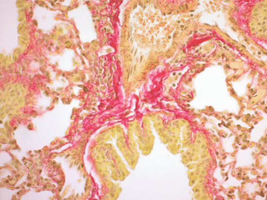
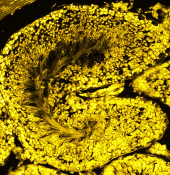
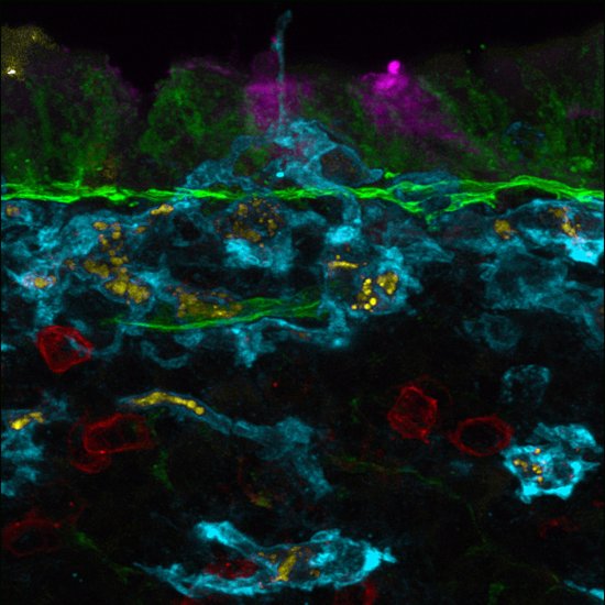
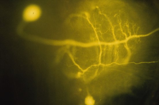
Montage issu d’une vidéo réalisée en microscopie confocale biphotonique (plateforme ImagoSeine, Institut Jacques Monod) illustrant le développement embryonnaire d’un nématode "Caenorhabditis elegans" exprimant un marqueur de membrane fluorescent. Les différents stades du développement sont répartis chronologiquement. Le volume cellulaire est progressivement réduit à chaque division cellulaire.
The use of media visible on the CNRS Images Platform can be granted on request. Any reproduction or representation is forbidden without prior authorization from CNRS Images (except for resources under Creative Commons license).
No modification of an image may be made without the prior consent of CNRS Images.
No use of an image for advertising purposes or distribution to a third party may be made without the prior agreement of CNRS Images.
For more information, please consult our general conditions
2019
Our work is guided by the way scientists question the world around them and we translate their research into images to help people to understand the world better and to awaken their curiosity and wonderment.