Production year
2011

© Hugues LELOUARD / CIML / INSERM / CNRS Images
20170103_0007
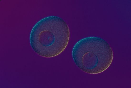
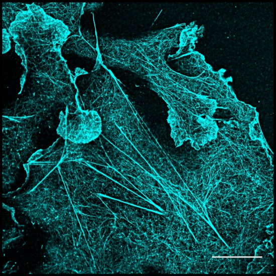
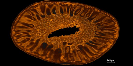
In a Peyer's patch (one of the constituents of the lymphoid tissue associated with the intestine) in a mouse, a dendritic cell expressing lysozyme (also known as LysoDC) sends a dendrite via a transcellular pore in an M cell to sense light inside the intestine, observed using confocal microscopy. Immunofluorescent marking shows the dendritic cells in cyan, the lysozyme in yellow, the M cells in magenta, the T lymphocytes in red, and lastly, the epithelial basement membrane and vessels in green.
The use of media visible on the CNRS Images Platform can be granted on request. Any reproduction or representation is forbidden without prior authorization from CNRS Images (except for resources under Creative Commons license).
No modification of an image may be made without the prior consent of CNRS Images.
No use of an image for advertising purposes or distribution to a third party may be made without the prior agreement of CNRS Images.
For more information, please consult our general conditions
2011
Our work is guided by the way scientists question the world around them and we translate their research into images to help people to understand the world better and to awaken their curiosity and wonderment.