Production year
2017

© Sébastien MAILFERT / Roxane FABRE / Yannick HAMON / CIML / INSERM / CNRS Images
20170103_0012
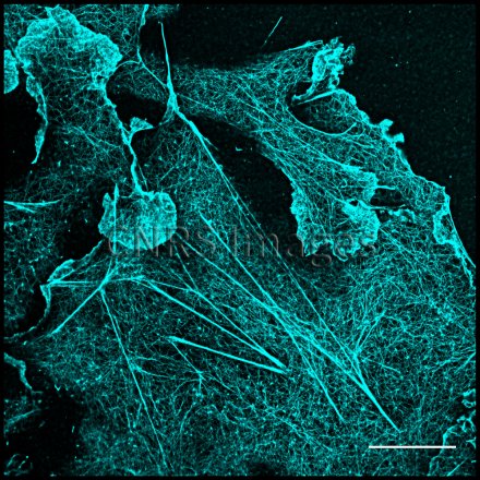
Cellules de fibroblastes rénaux de singe vert africain COS-7 observées en microscopie confocale à fluorescence super-résolution dSTORM (direct stochastic optical reconstruction microscopy). Les cellules sont marquées avec la phalloïdine conjuguée à Alexa Fluor 647. On observe ainsi les fibres de stress et les filaments d'actine avec une résolution de 20 nm. La technique de super résolution, ici le dSTORM permet d’observer des structures par fluorescence de plus en plus finement. Ici, les filaments d'actine sont clairement définis, permettant ainsi d'étudier au mieux leur rôle dans certains processus cellulaires vitaux, tels que l’adhésion ou la mobilité cellulaire, le transport intracellulaire et la morphogenèse.
The use of media visible on the CNRS Images Platform can be granted on request. Any reproduction or representation is forbidden without prior authorization from CNRS Images (except for resources under Creative Commons license).
No modification of an image may be made without the prior consent of CNRS Images.
No use of an image for advertising purposes or distribution to a third party may be made without the prior agreement of CNRS Images.
For more information, please consult our general conditions
2017
Our work is guided by the way scientists question the world around them and we translate their research into images to help people to understand the world better and to awaken their curiosity and wonderment.