Production year
2013

© Cyril FRESILLON/CNRS Images
20130001_0233
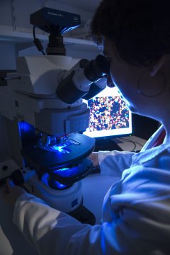
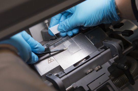
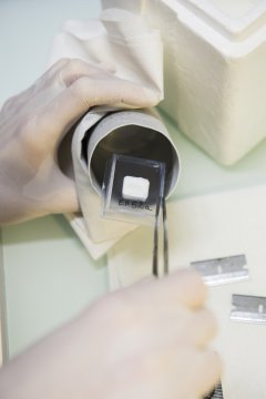
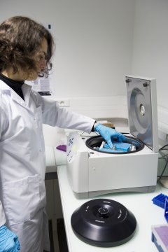
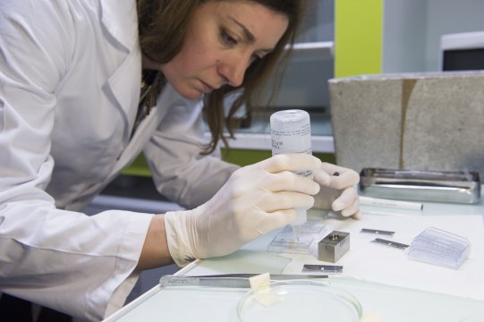
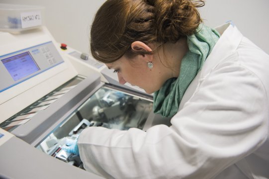
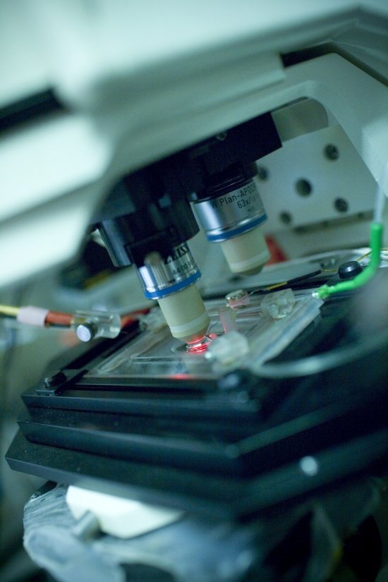
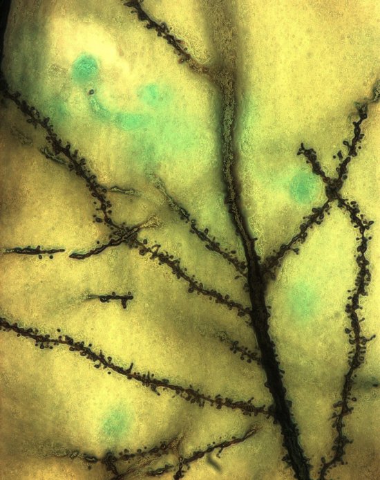
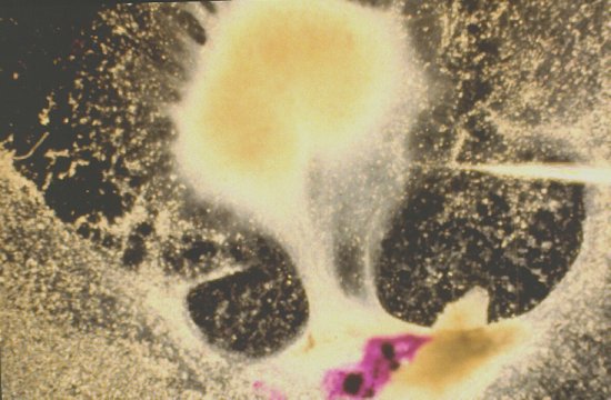
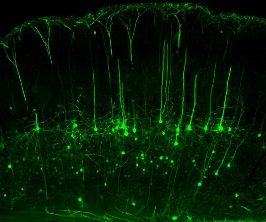
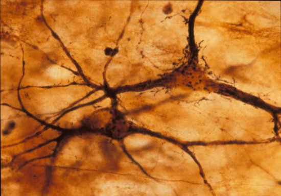
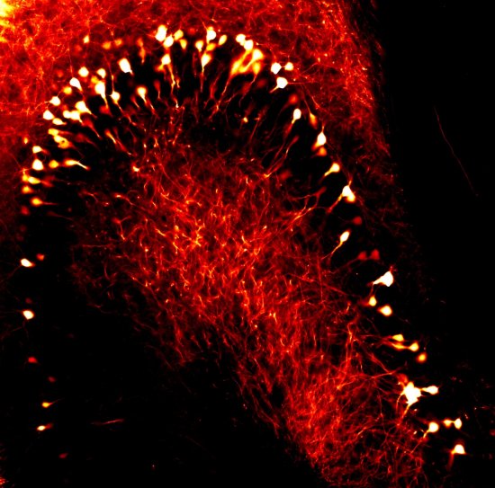
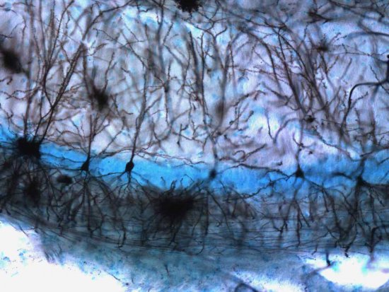
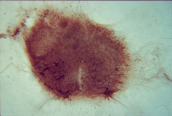
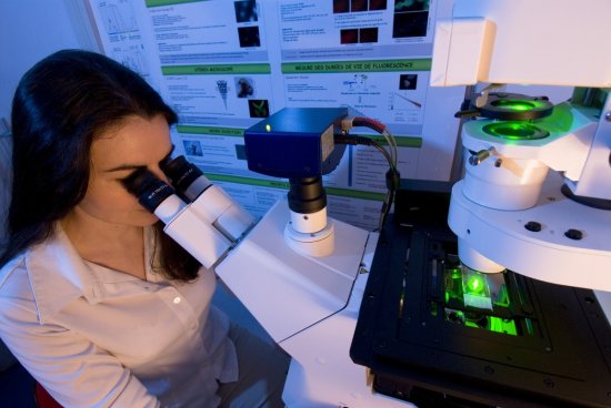
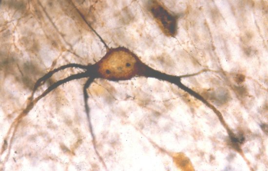
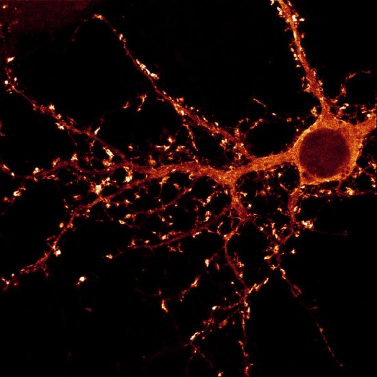
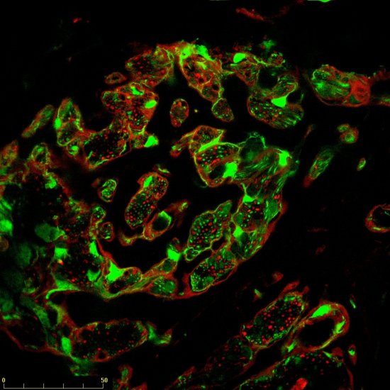
Observation et acquisition d'images de cellules neurales de souris, au microscope à fluorescence. Ces cellules qui composent le tissu nerveux sont de trois types : les neurones, les astrocytes et les oligodendrocytes. Les astrocytes (en rouge) ont été colorés grâce au marqueur GFAP (Glial fibrillary acidic protein). Les noyaux des cellules sont colorés en bleu.
The use of media visible on the CNRS Images Platform can be granted on request. Any reproduction or representation is forbidden without prior authorization from CNRS Images (except for resources under Creative Commons license).
No modification of an image may be made without the prior consent of CNRS Images.
No use of an image for advertising purposes or distribution to a third party may be made without the prior agreement of CNRS Images.
For more information, please consult our general conditions
2013
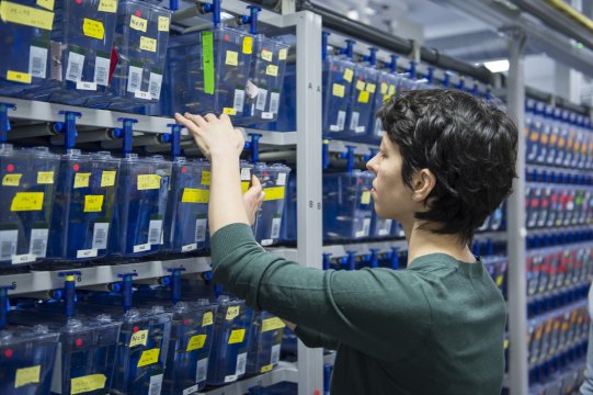
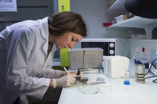
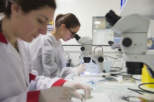
Our work is guided by the way scientists question the world around them and we translate their research into images to help people to understand the world better and to awaken their curiosity and wonderment.