Production year
2017

© Jean-Claude MOSCHETTI / IRCER / CNRS Images
20170068_0057
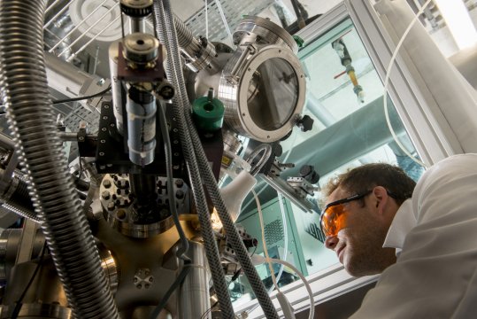
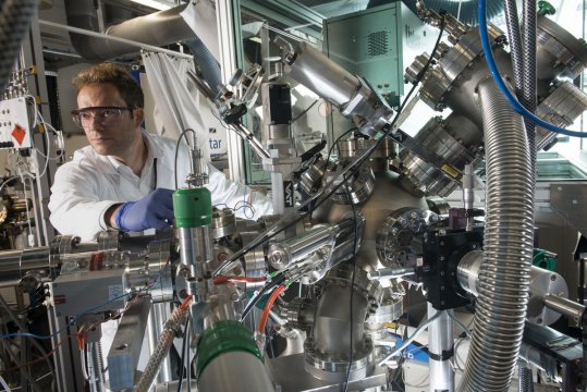
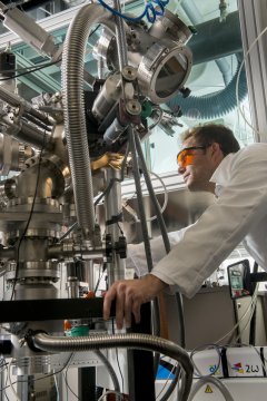
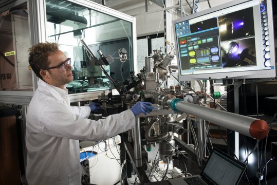
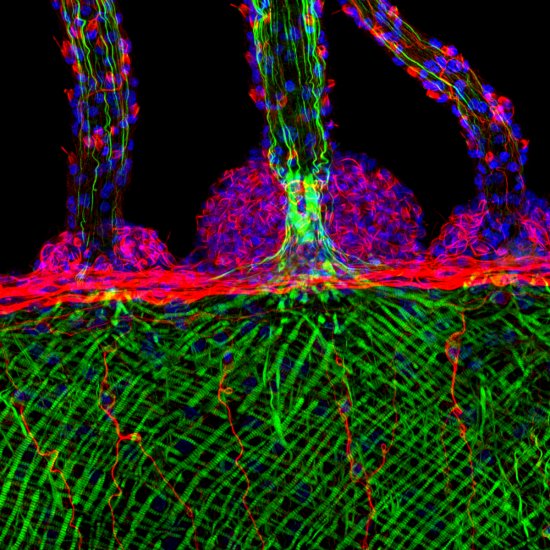
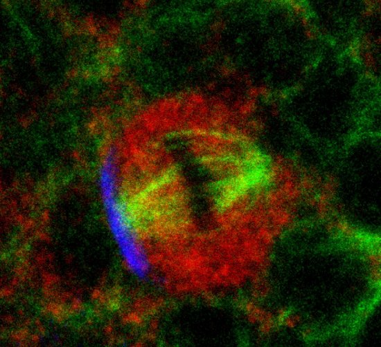
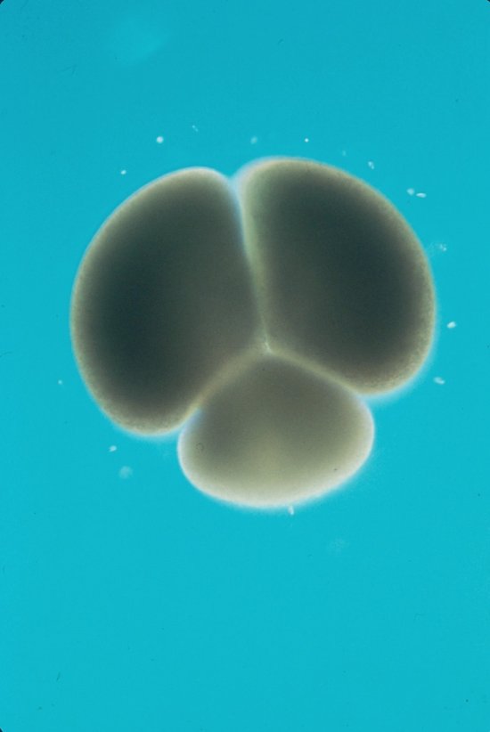
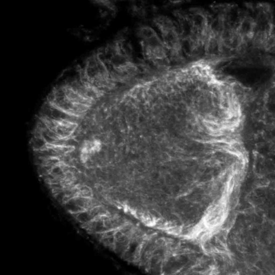
Evaluation of the adhesion of murine pre-osteoblast cells to a ceramic material (hydroxyapatite) using immunofluorescence labelling. This involves observing the morphology of the cells and the way in which they have adhered to the surface of the ceramic. These cells are analysed by viewing the structures or markers detected via indirect coupling to various fluorescent molecules. The cell nuclei can be seen in the bottom left of the screen. In the top left, the detection of vinculin allows the points of adhesion to be viewed. In the top right, it is possible to see a constituent of the cytoskeleton, actin, which is associated with vinculin in the cell adhesion areas, and which also indicates the morphology of the cell. Finally, the three images are superimposed upon one another in the bottom right corner. This research is performed as part of efforts to evaluate the biological properties of optimised ceramic materials destined for applications in bone tissue engineering (bone substitutes).
The use of media visible on the CNRS Images Platform can be granted on request. Any reproduction or representation is forbidden without prior authorization from CNRS Images (except for resources under Creative Commons license).
No modification of an image may be made without the prior consent of CNRS Images.
No use of an image for advertising purposes or distribution to a third party may be made without the prior agreement of CNRS Images.
For more information, please consult our general conditions
2017
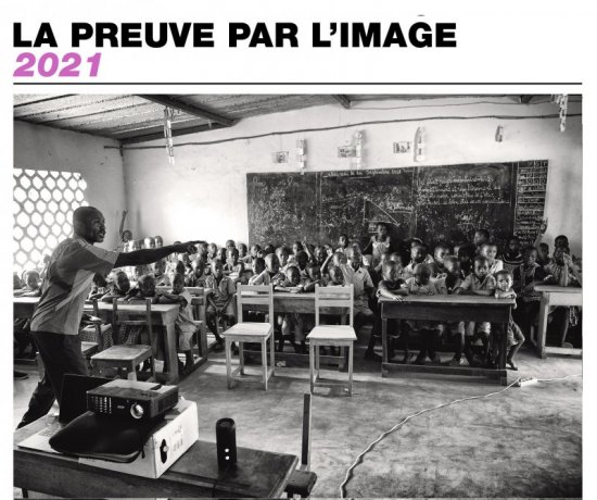
Our work is guided by the way scientists question the world around them and we translate their research into images to help people to understand the world better and to awaken their curiosity and wonderment.