Production year
2016

© Serge VAN DE PAVERT / Carole SIRET / CIML / CNRS Images
20170103_0017
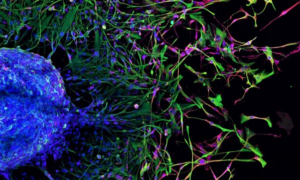
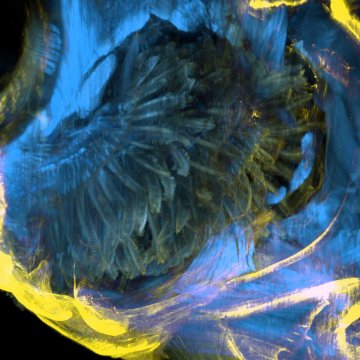
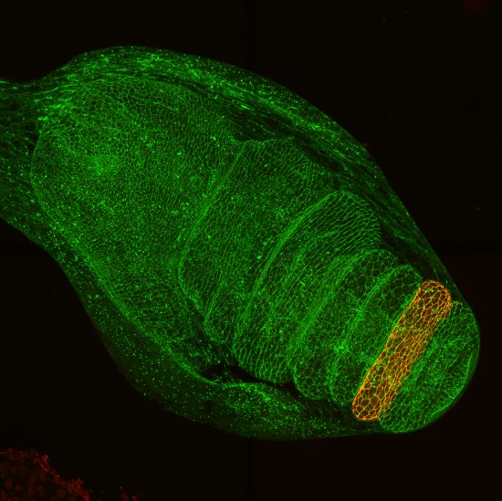
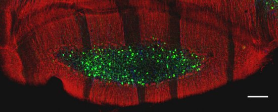
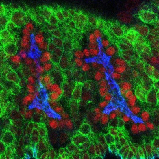
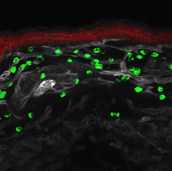
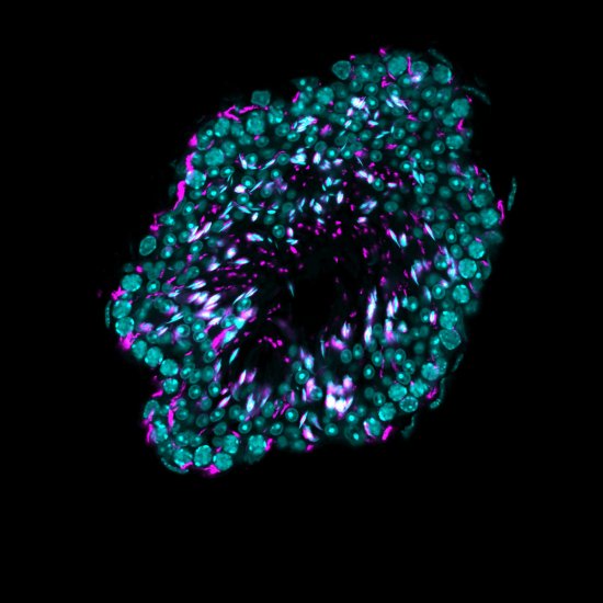
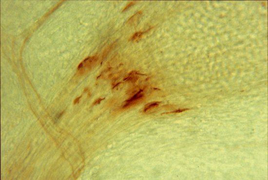
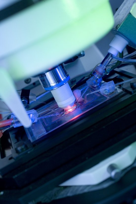
The embryonic development of lymph nodes, small organs that are essential for the immune response, is now well understood. Using light-sheet microscopy, scientists were able to determinethe processes at work in this 13.5-day-old mouse embryo. Shown in blue are the lymphoid cells (LTi), derived from the haemogenic endothelium, a tissue specific to the embryo. They move into the liver where they multiply and then migrate to the rest of the body, where they give rise to the lymph nodes. The 3D information obtained is used to track the interactions of the lymph nodes with their environment, in particular with nerve cells, in green, and blood vessels, in white. Lymphatic endothelial cells and some macrophages are displayed in red. This image was taken using light sheet microscopy. It is one of the winners of the 2022 La preuve par l’image (LPPI) photo competition.
The use of media visible on the CNRS Images Platform can be granted on request. Any reproduction or representation is forbidden without prior authorization from CNRS Images (except for resources under Creative Commons license).
No modification of an image may be made without the prior consent of CNRS Images.
No use of an image for advertising purposes or distribution to a third party may be made without the prior agreement of CNRS Images.
For more information, please consult our general conditions
2016
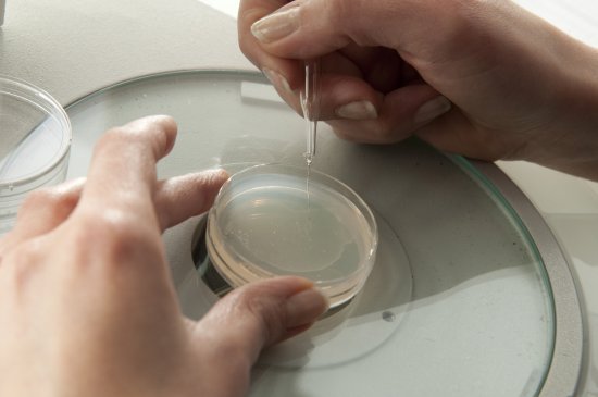
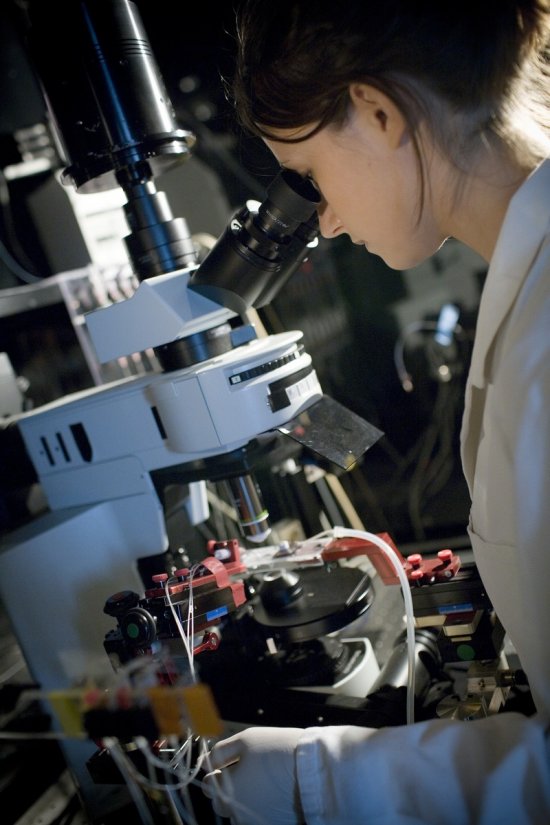
Our work is guided by the way scientists question the world around them and we translate their research into images to help people to understand the world better and to awaken their curiosity and wonderment.