Production year
2014

© Sébastien MAILFERT / Noushin MOSSADEGH / CIML / INSERM / AMU / CNRS Images
20170103_0011
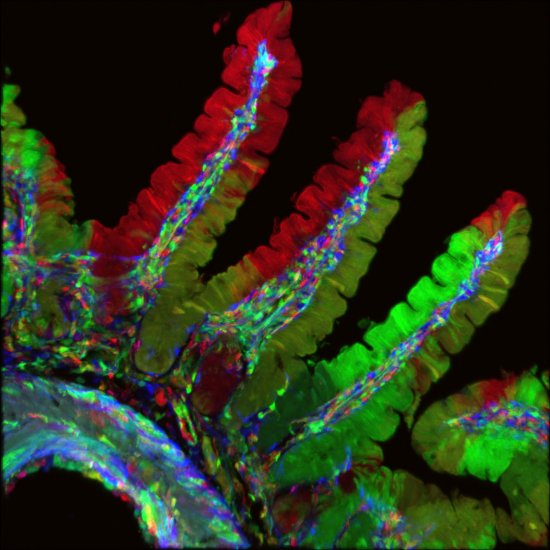
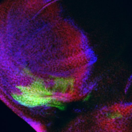
A 20 µm thick cross-section of a seminiferous tubule in a mouse testicle, observed using multicolour confocal microscopy. The heads of the sperm cells have been marked in red by immunofluorescence (phalloidin marked with the fluorochrome Alexa-647), and their tails are shown in cyan (DAPI). Testicular macrophages are mobilised to defend sperm cells. By releasing specific molecules, these guardians of fertility prevent other immune system agents from entering the testes. A recent study described two types of testicular macrophage, establishing the origin, development and characteristics of these immune cells. The first population of testicular macrophage are embryonic in nature. Peritubular macrophages, on the other hand, are found around the seminiferous tubules. Using a new cell tracing method, researchers have successfully tracked peritubular macrophages (which travel from bone marrow to the testes), which in mice do not appear until two weeks after birth, corresponding to puberty in humans. Surprisingly, once established in the testes, both macrophage populations remain there for the rest of their lives. This discovery may enable the development of innovative male infertility treatments. This image was obtained using confocal microscopy which uses laser beams to scan the specimen, point by point. This technique can be used to produce thin optical cross-sections that reveal the locations of numerous entities and cell types (T and B lymphocytes, dendrites, macrophages, actins, nuclei, etc.) in a tissue slice. This enables scientists to compare the locations and numbers of entities in various circumstances, such as in patients suffering from diseases, for example, in order to shed light on the workings of the immune system. This technique is now widely used in laboratories, and is constantly being improved in terms of its resolution and sensitivity, as well as the possible colours and contrasts.
The use of media visible on the CNRS Images Platform can be granted on request. Any reproduction or representation is forbidden without prior authorization from CNRS Images (except for resources under Creative Commons license).
No modification of an image may be made without the prior consent of CNRS Images.
No use of an image for advertising purposes or distribution to a third party may be made without the prior agreement of CNRS Images.
For more information, please consult our general conditions
2014
Our work is guided by the way scientists question the world around them and we translate their research into images to help people to understand the world better and to awaken their curiosity and wonderment.