© Olivia BERTHOUMIEU / LCC / CNRS Images
Reference
20160084_0001
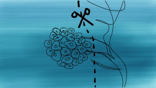
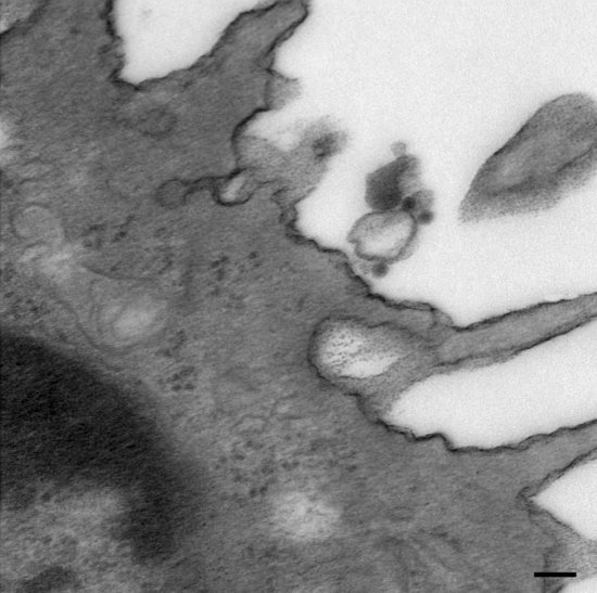
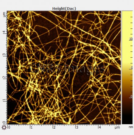
Stages in the formation of the amyloid fibres encountered in Alzheimer’s disease
Images made by atomic force microscopy (AFM) and transmission electronic microscopy (TEM) showing stages in the formation of the amyloid fibres encountered in Alzheimer’s disease. From small protein fragments, fibrils build and lengthen to produce the fibres (visible in the image) that are found in the tangled senile plaques detected in Alzheimer’s patients.
The use of media visible on the CNRS Images Platform can be granted on request. Any reproduction or representation is forbidden without prior authorization from CNRS Images (except for resources under Creative Commons license).
No modification of an image may be made without the prior consent of CNRS Images.
No use of an image for advertising purposes or distribution to a third party may be made without the prior agreement of CNRS Images.
For more information, please consult our general conditions

