© CNRS Images/media 2001
Reference
910
First 3-D picture of a prion (The)
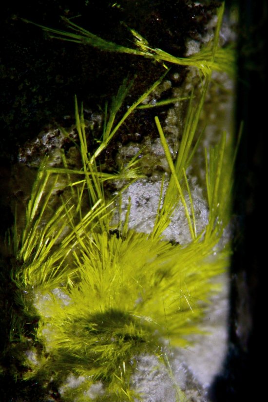
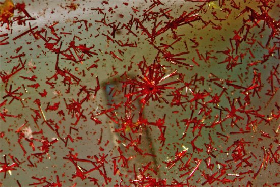
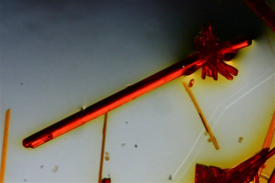
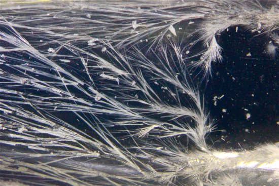
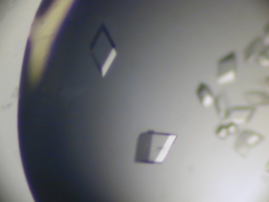
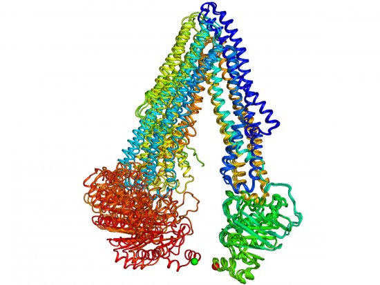
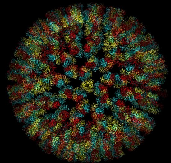
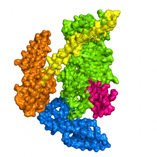
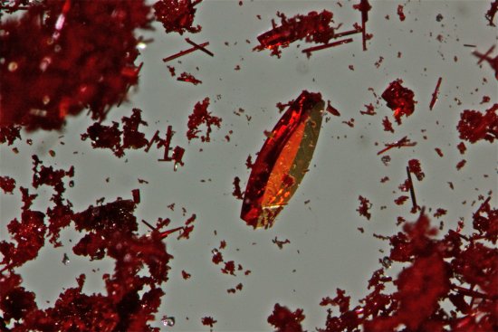
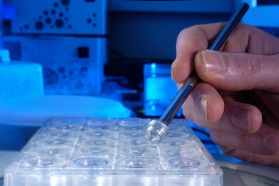
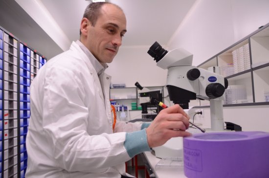
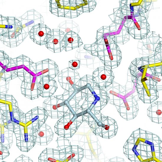
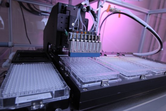
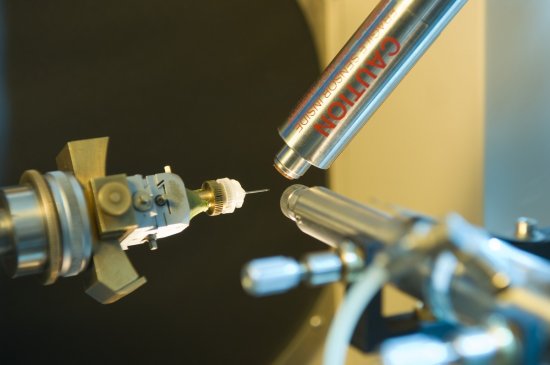
Mad cow disease, like other neurodegenerative diseases, is probably due to the presence of an abnormal form of the prion, a protein found in mammalian nerve cells. A collaborative project by a team of biochemists from the Laboratoire d'enzymologie et biochimie structurale in Gif-sur-Yvette and a team of physicists from the ESRF (European Synchrotron Radiation Facility) in Grenoble has led to an understanding of the three-dimensional structure of the yeast prion.The prion crystals are positioned in the path of a beam of x-rays produced in the Grenoble synchrotron. The x-rays strike the crystals, and rays diffracted in different directions form spots on a detector. Based on the position and relative intensity of the spots, the three-dimensional structure of the prion protein can be reconstructed and modeled by computer. The researchers have observed the existence of a flexible zone inside the molecule which may be the source of the appearance and transmission of the abnormal prion.
Duration
Production year
Définition
Color
Sound
Version(s)
Original material
The use of media visible on the CNRS Images Platform can be granted on request. Any reproduction or representation is forbidden without prior authorization from CNRS Images (except for resources under Creative Commons license).
No modification of an image may be made without the prior consent of CNRS Images.
No use of an image for advertising purposes or distribution to a third party may be made without the prior agreement of CNRS Images.
For more information, please consult our general conditions
