Production year
2006

© François JANNIN/CNRS Images
20060001_1165
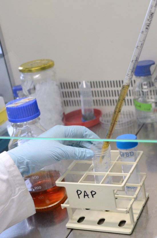
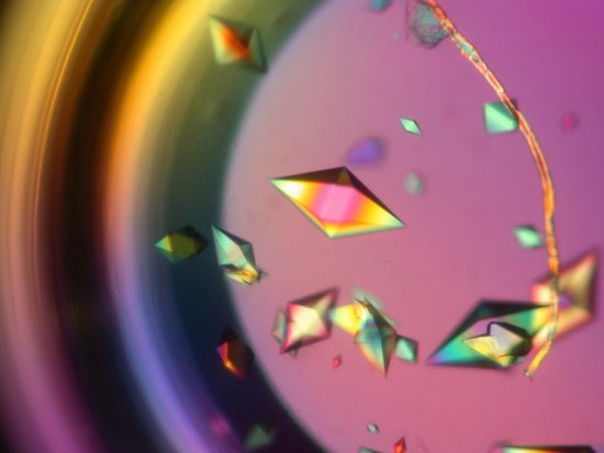
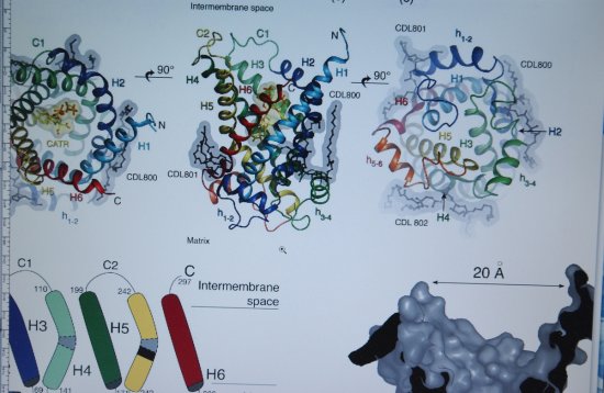
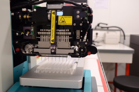
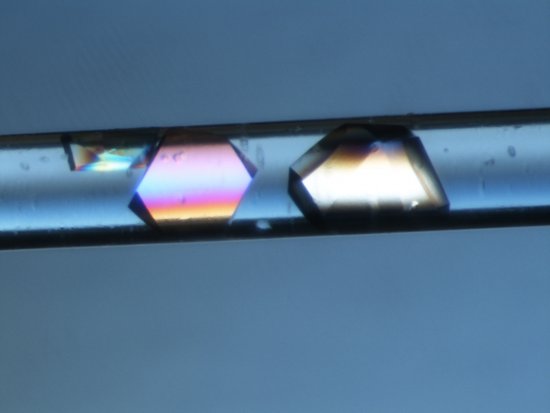
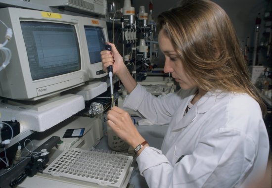
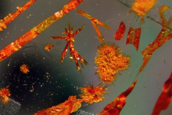
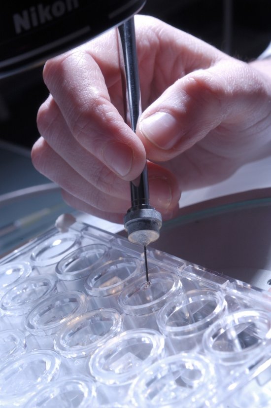
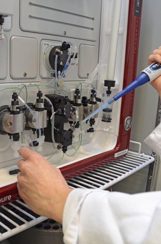
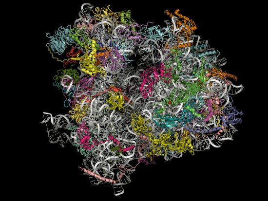
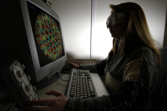
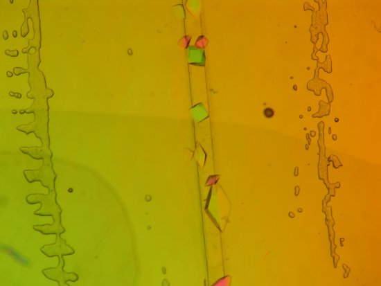
Détail du diffractomètre pour l'analyse cristallographique aux rayons X. Le cristal congelé est fixé à l'extrémité de la petite tige à gauche. Un flux d'azote maintenant le cristal à 100 kelvin arrive par le tuyau en haut. Détermination de la structure 3D de macromolécules biologiques.
The use of media visible on the CNRS Images Platform can be granted on request. Any reproduction or representation is forbidden without prior authorization from CNRS Images (except for resources under Creative Commons license).
No modification of an image may be made without the prior consent of CNRS Images.
No use of an image for advertising purposes or distribution to a third party may be made without the prior agreement of CNRS Images.
For more information, please consult our general conditions
2006
Our work is guided by the way scientists question the world around them and we translate their research into images to help people to understand the world better and to awaken their curiosity and wonderment.