Production year
2021

© Louise GRIVEAU / Emilie CHRISTIN / LBTI / INMG / CNRS Images
20210137_0012
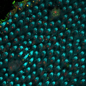
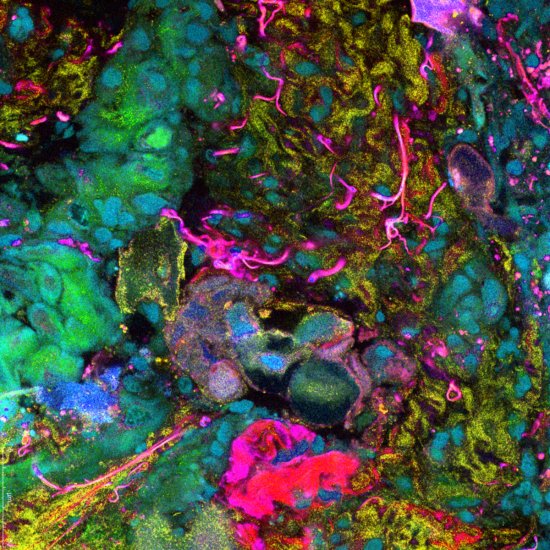
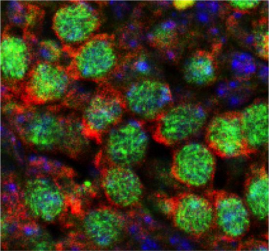
When muscle precursor cells fuse, they happen to form the swirling patterns shown here, unmistakably reminiscent of Van Gogh’s painting, The Starry Night. In these microscopic whorls, their cell nuclei can be seen in cyan, the actin cytoskeleton in blue, and, in yellow, a protein indicating the formation of new muscle fibres. The cells are grown on an ultra-nutritious medium – Matrigel – that serves as a support for them. Their behaviour is subsequently studied with the aim of developing innovative biomaterials, which can be used as dressings to fill the wounds left by deep tissue injuries. The challenge is to find new therapeutic solutions for the management of chronic or complex lesions, such as diabetic wounds and bullet wounds, and for the improvement of symptoms related to muscular dystrophy. This image is a winner of the 2021 La preuve par l'image (LPPI) competition.
The use of media visible on the CNRS Images Platform can be granted on request. Any reproduction or representation is forbidden without prior authorization from CNRS Images (except for resources under Creative Commons license).
No modification of an image may be made without the prior consent of CNRS Images.
No use of an image for advertising purposes or distribution to a third party may be made without the prior agreement of CNRS Images.
For more information, please consult our general conditions
2021
Our work is guided by the way scientists question the world around them and we translate their research into images to help people to understand the world better and to awaken their curiosity and wonderment.