Production year
2020

© Louise SIMONNET / CIML / CNRS Images
20210137_0008
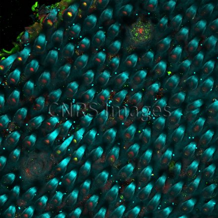
The tongue is covered with taste buds that can perceive five tastes: sweet, salty, sour, bitter and umami. This organ remains poorly understood, both regarding its structure and the different cells that make it up. However, immunofluorescence provides a better view of it. The image shows the surface of an epithelium (a tissue that has a covering function) of a rodent’s tongue. The small spikes are filiform papillae (blue autofluorescence), which play a mechanical role in bringing food to the back of the mouth, while the two circular galaxy-like areas (in green and red), are taste buds. Improving existing knowledge of the cellular composition of the tongue will make it possible to understand what happens in the event of an infection or a loss of taste. This image is a winner of the 2021 La preuve par l'image (LPPI) competition.
The use of media visible on the CNRS Images Platform can be granted on request. Any reproduction or representation is forbidden without prior authorization from CNRS Images (except for resources under Creative Commons license).
No modification of an image may be made without the prior consent of CNRS Images.
No use of an image for advertising purposes or distribution to a third party may be made without the prior agreement of CNRS Images.
For more information, please consult our general conditions
2020
Our work is guided by the way scientists question the world around them and we translate their research into images to help people to understand the world better and to awaken their curiosity and wonderment.