© Anne-Catherine SCHMIT / IBMP / CNRS Images
Reference
20170094_0002
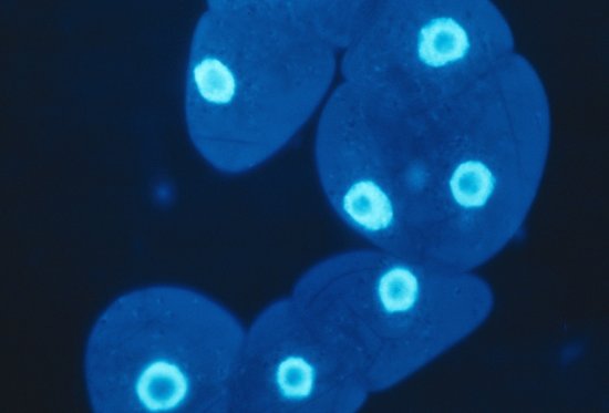
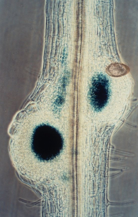
Marquage des centromères dans une pointe de racine d’Arabette des dames
Marking centromeres in a root tip of thale cress (arabidopsis thaliana). This line overexpresses a centromeric histone H3 variant (YFP-CENH3), which can be observed in the nuclei, where it appears in blue. The protein GIP1-GFP (shown in green) colocalises with YFP-CENH3 and is also distributes in the mitotic spindle of dividing cells. Propidium iodide (shown in red) reveals the walls of the living cells. The aim is to characterise the molecular functions of GIP proteins, which are present in almost all eukaryote and multi-functional organisms. They play a key role in microtubule formation and in the robustness of the spindle apparatus. In severe knock-down mutants, the loss of expression of these proteins leads to chromosome cohesion defects. This cohesion is measured by the distance between two centromeres of a replicated chromosome. To test the link between GIP proteins and centromeres, scientists successfully used a co-immunoprecipitation assay to prove their joint presence in protein complexes in vivo, and their colocalisation in situ, as illustrated in this image. This research is being pursued, with the aim of understanding the role of GIP in addressing, maintenance and regulation of the proteins with a role in centromere identity.
Add to my selection
Terms of use
The use of media visible on the CNRS Images Platform can be granted on request. Any reproduction or representation is forbidden without prior authorization from CNRS Images (except for resources under Creative Commons license).
No modification of an image may be made without the prior consent of CNRS Images.
No use of an image for advertising purposes or distribution to a third party may be made without the prior agreement of CNRS Images.
For more information, please consult our general conditions