© Aude CERUTTI / Alain JAUNEAU / Laurent NOEL / LIPM / TRI / CNRS Images
Reference
20170089_0004
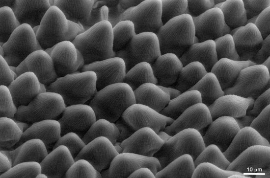
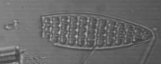
Chou-fleur infecté avec la bactérie phytopathogène "Xanthomonas campestris"
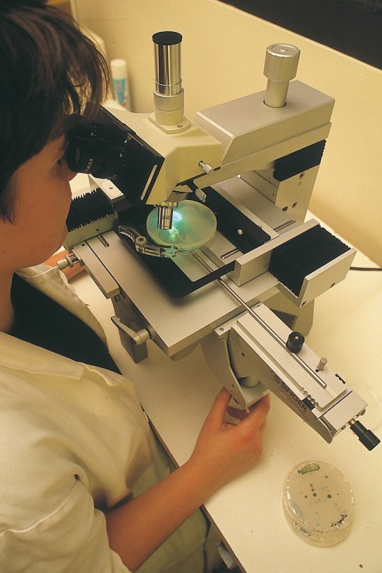
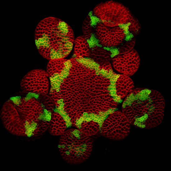
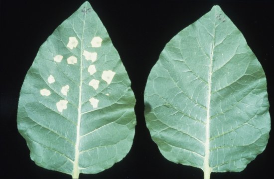
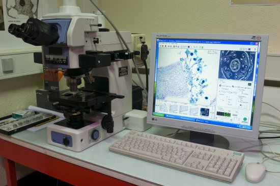
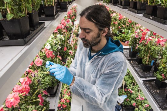
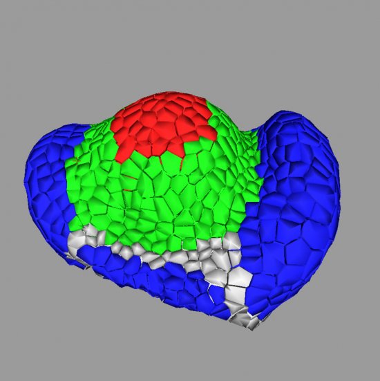
Surface of a cauliflower hydathode infected with the phytopathogenic bacterium Xanthomonas campestris, observed using a cryo-scanning electron microscope. The images have been manually coloured using the Gimp2 software package. The leaf surface is shown in green. A hydathode pore is visible. The bacteria can be seen in orange. These examinations were carried out as part of research to obtain a full understanding of the mechanisms through which plants (cauliflower or Arabidopsis thaliana) are infected by the pathogenic bacterium Xanthomonas campestris. The use of cryo-preparation with scanning electron microscopy makes it possible to keep the biological object in its original state (frozen, hydrated), reducing the potential artefacts associated with the chemical preparation of samples. In this way, the detailed anatomy of structures has been described for various plant species, whether infected or not by Xanthomonas. Initial results have helped clarify the structure of the hydathodes located on the edge of the leaves, and demonstrated that the hydathode pores (which resemble stomata) are the points at which the Xanthomonas bacterium infects the leaves. Based on these conclusions, further research is being done into controlling infection at a molecular level.
Add to my selection
Terms of use
The use of media visible on the CNRS Images Platform can be granted on request. Any reproduction or representation is forbidden without prior authorization from CNRS Images (except for resources under Creative Commons license).
No modification of an image may be made without the prior consent of CNRS Images.
No use of an image for advertising purposes or distribution to a third party may be made without the prior agreement of CNRS Images.
For more information, please consult our general conditions