Production year
2017
Attention, non CNRS staff

© Christophe HARGOUES / IGMM / CNRS Images
20170073_0048
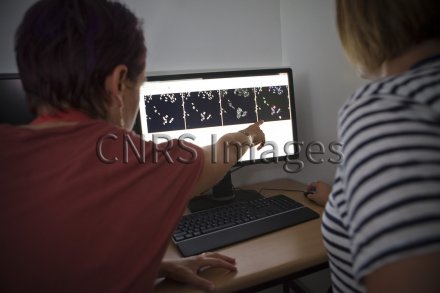
Observation of microscope images of cell lines from a human carcinoma produced using an automatic inverted microscope, which allows high-speed image acquisition. The first three images correspond to the acquisition of each colour on a different channel, and the fourth is a reconstruction of the superimposed images with artificial colouring: the nucleus in blue, the protein of interest in green and the cytoplasm in red. These research scientists are interested in certain proteins with a view to understanding how they work on cell lines used as easily observable models. Afterwards, they will be able to transfer these observations to examine how the proteins interact with HIV (human immunodeficiency virus).
The use of media visible on the CNRS Images Platform can be granted on request. Any reproduction or representation is forbidden without prior authorization from CNRS Images (except for resources under Creative Commons license).
No modification of an image may be made without the prior consent of CNRS Images.
No use of an image for advertising purposes or distribution to a third party may be made without the prior agreement of CNRS Images.
For more information, please consult our general conditions
2017
Our work is guided by the way scientists question the world around them and we translate their research into images to help people to understand the world better and to awaken their curiosity and wonderment.