© Christophe LETERRIER/NICN/CNRS Images
Reference
20170024_0001
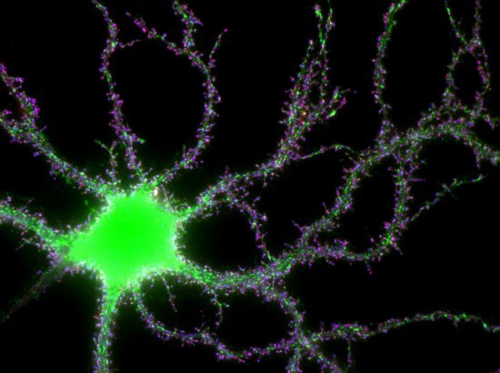
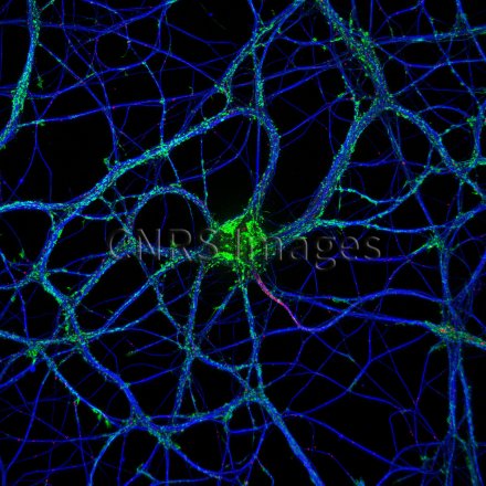
Microarchitecture d'un neurone en culture
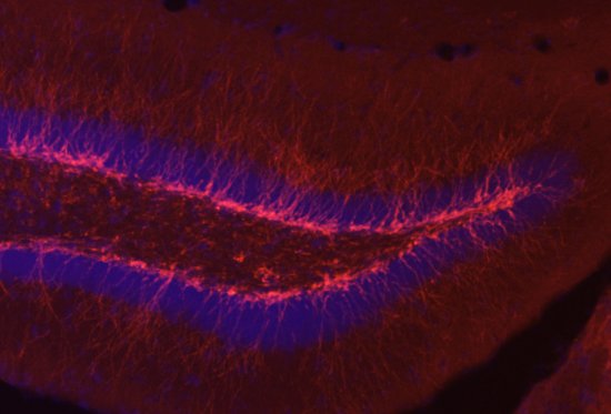
Microarchitecture of a three-week-old neuron in culture, observed using fluorescence microscopy. This neuron was taken from a rat embryo hippocampus. The hippocampus is a brain structure involved in learning and memory processes. Two components of the cytoskeleton, microtubules are marked in blue and actin filaments in green. The cytoskeleton is essential for the proper functioning of the neuron. It enables it to acquire and retain its shape, as well as organise the transport of proteins to the various cell compartments (axon, dendrites, synapses, etc.). In red, neurofascin protein is concentrated in way of the initial axon segment. This segment is the starting point of the electrical signal, known as the action potential, which propagates along the axon towards the other neurons.
The use of media visible on the CNRS Images Platform can be granted on request. Any reproduction or representation is forbidden without prior authorization from CNRS Images (except for resources under Creative Commons license).
No modification of an image may be made without the prior consent of CNRS Images.
No use of an image for advertising purposes or distribution to a third party may be made without the prior agreement of CNRS Images.
For more information, please consult our general conditions

