© Didier MONTARRAS/Institut Pasteur/CNRS Images
Reference
20130001_1640
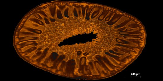
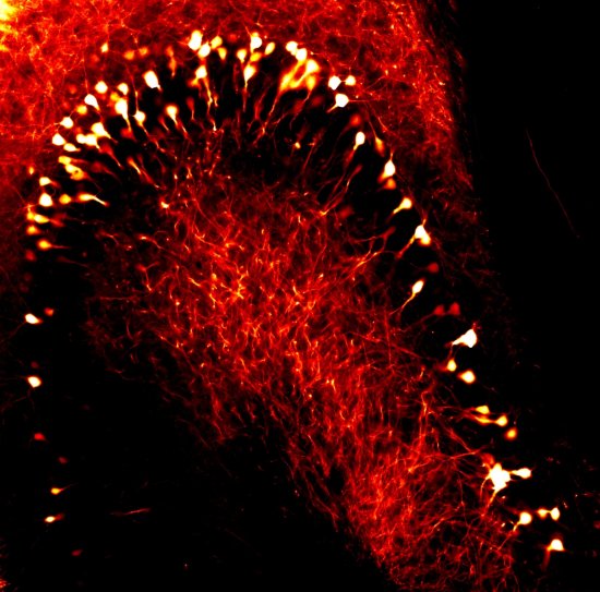
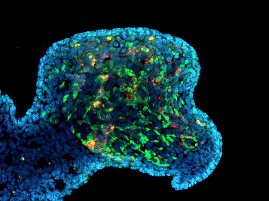
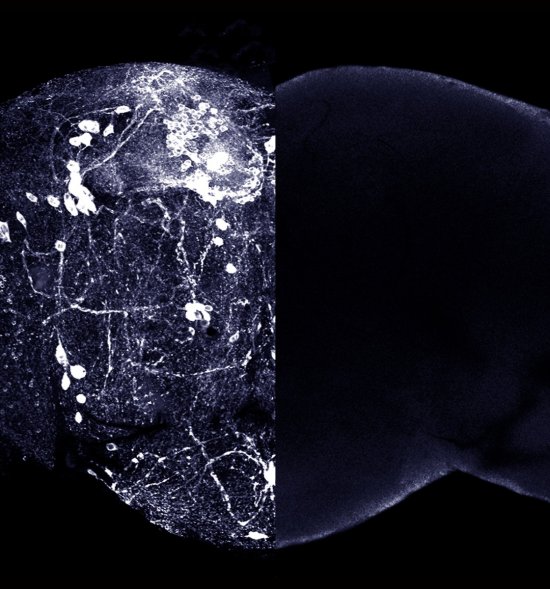
Transverse sections of mouse skeletal muscles
Transverse sections of mouse skeletal muscles, with muscle fibers in red and their nuclei in blue. At the top, a non-injured muscle. The nuclei of the fibers are found at the periphery. The photograph at the bottom shows a regenerating muscle, 12 days after injury. Regenerating fibers have centrally located nuclei and many of them are smaller in size. The capacity of skeletal muscles to undergo regeneration depends on skeletal muscle stem cells, called myosatellite cells. Upon muscle injury, myosatellite cells undergo activation, proliferate, differentiate and fuse with each other to form new fibers.
Add to my selection
Terms of use
The use of media visible on the CNRS Images Platform can be granted on request. Any reproduction or representation is forbidden without prior authorization from CNRS Images (except for resources under Creative Commons license).
No modification of an image may be made without the prior consent of CNRS Images.
No use of an image for advertising purposes or distribution to a third party may be made without the prior agreement of CNRS Images.
For more information, please consult our general conditions