Production year
2004

© Emmanuel PERRIN/CNRS Images
20040001_0086
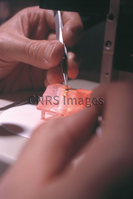
Préparation d'un cerveau d'abeille pour l'imagerie calcique in vivo. L'abeille est placée dans une chambre en plexiglas, et des marquages de son cerveau sont réalisés avec une sonde fluorescente spécifique du calcium, ce qui permet de suivre l'activité des structures nerveuses au cours du temps, en mesurant les variations intracellulaires en ions calcium déclenchées par divers stimuli, en particulier olfactifs.
The use of media visible on the CNRS Images Platform can be granted on request. Any reproduction or representation is forbidden without prior authorization from CNRS Images (except for resources under Creative Commons license).
No modification of an image may be made without the prior consent of CNRS Images.
No use of an image for advertising purposes or distribution to a third party may be made without the prior agreement of CNRS Images.
For more information, please consult our general conditions
2004
Our work is guided by the way scientists question the world around them and we translate their research into images to help people to understand the world better and to awaken their curiosity and wonderment.