© Thomas DENEUX / UNIC / CNRS Images
Reference
20170057_0001
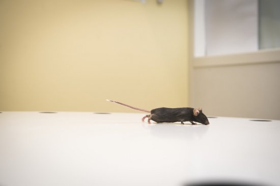
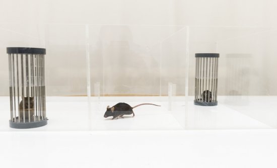
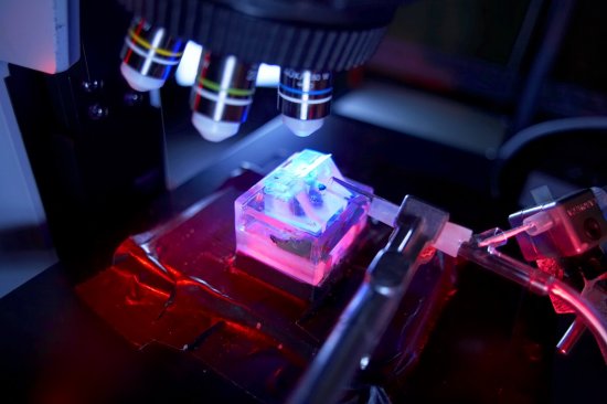
Neurones du cortex sensoriel d’une souris éveillée observés par microscopie de fluorescence
Sensory cortex neurons in an awake mouse viewed using a two-photon excitation fluorescence microscope. The neurons have been transfected with an adeno-associated virus (AAV) used for gene transfer, activating a fluorescent protein sensitive to calcium. The variations in fluorescence over time provide information about the electrical activity of the neurons. The awake mouse undergoes sensory stimulation, potentially learning to respond to it according to a pre-established behavioural protocol. Analysing the activity of neurons helps us understand how the neural network processes sensory information.
Add to my selection
Terms of use
The use of media visible on the CNRS Images Platform can be granted on request. Any reproduction or representation is forbidden without prior authorization from CNRS Images (except for resources under Creative Commons license).
No modification of an image may be made without the prior consent of CNRS Images.
No use of an image for advertising purposes or distribution to a third party may be made without the prior agreement of CNRS Images.
For more information, please consult our general conditions