© CNRS Images media - 2001
Reference
945
Using the tools of physics in biology and medicine
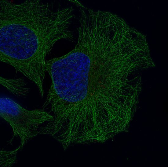
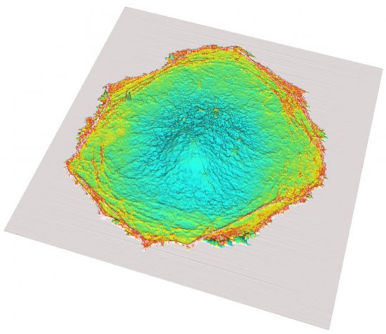
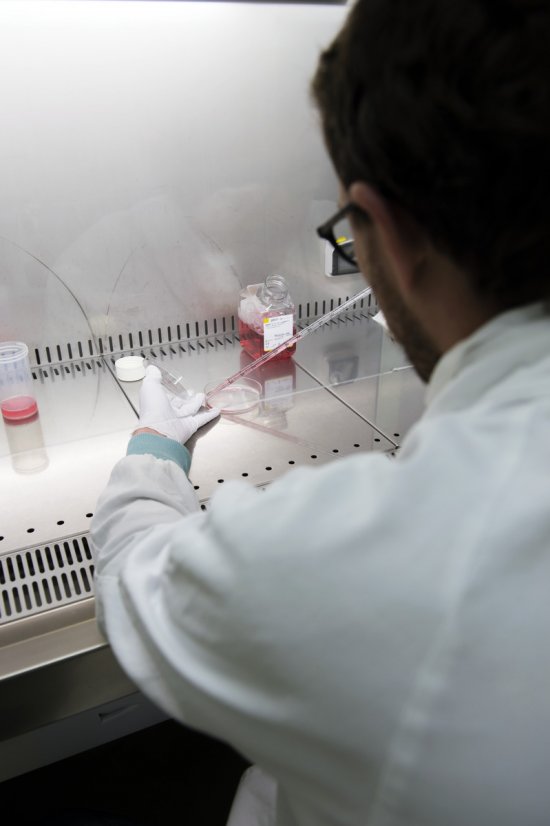
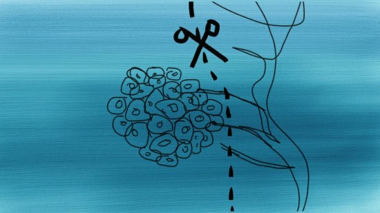
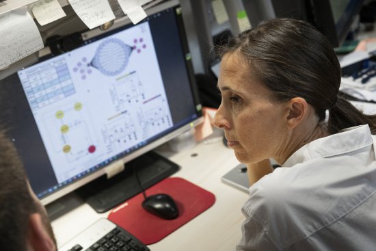
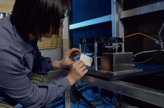
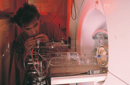
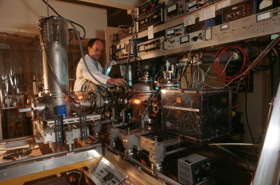
Seven researchers present the medical and biological applications of techniques originally developed in the field of physics.Sigrid Avrillier (Laboratoire de physique des lasers, Villetaneuse) describes a laser sensor which can be used to measure the oxygenation properties of tissues.Philippe Lanièce (Institut de physique nucléaire, Orsay) describes a high-resolution tomography system (TOHR) used to obtain an image of the brain of a small animal.Pascal Laugier (Laboratoire d'imagerie paramétrique, Faculty of medicine, Paris) investigates the use of ultrasound in medical imaging.Bernard Renault (Laboratoire Neurosciences cognitives et imagerie cérébrale, Pitié Salpétrière hospital, Paris) uses magneto-encephalography and electro-encephalography to localize the brain's areas of activity and connect them to a patient's gestures. Mathias Fink (Laboratoire Ondes et acoustique, Ecole supérieure de physique et de chimie industrielle, Paris) presents a very high frequency ultrasound detector which can visualize shear waves in tissue and differentiate them by texture (by identifying nodules harder than the soft tissue which surrounds them). Jacques Bittoun (Unité de recherche en résonance magnétique médicale, Kremlin Bicêtre hospital) uses MRI to study cardiac and pulmonary function. Didier Chatenay (Laboratoire de dynamique des fluides complexes, Strasbourg) is interested in the structure of RNA manipulated via laser in order to measure folding during transcription.
Duration
00:25:00
Production year
Définition
SD
Color
Color
Sound
Sound
Version(s)
French
Original material
Video DV Cam
Add to my selection
Terms of use
The use of media visible on the CNRS Images Platform can be granted on request. Any reproduction or representation is forbidden without prior authorization from CNRS Images (except for resources under Creative Commons license).
No modification of an image may be made without the prior consent of CNRS Images.
No use of an image for advertising purposes or distribution to a third party may be made without the prior agreement of CNRS Images.
For more information, please consult our general conditions