Production year
2023
Use only in the context of the LPPI competition

© Romina D'ANGELO / SigDYN / CRCT / IUCT-O / CNRS Images
20230104_0012
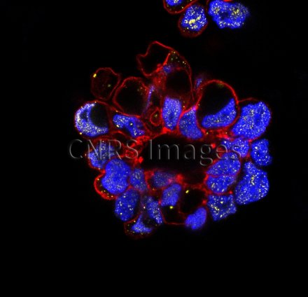
The visual appeal of this chromatic composition is deceptive. This is in fact a cluster of cancer cells from the peritoneal fluid of a patient with advanced ovarian cancer. This high-resolution image, which shows in detail the morphology of a cell cluster, reveals its metastatic potential. Under such circumstances, some nuclei, in blue, press up against the cell walls, in red. The yellow spots dotted around the interior of these same cells reveal the presence of a protein involved in the process of metastasis. Visual characterisation of cell clusters, which are both quickly and easily collected, could help improve the diagnosis and treatment of ovarian cancers.
The use of media visible on the CNRS Images Platform can be granted on request. Any reproduction or representation is forbidden without prior authorization from CNRS Images (except for resources under Creative Commons license).
No modification of an image may be made without the prior consent of CNRS Images.
No use of an image for advertising purposes or distribution to a third party may be made without the prior agreement of CNRS Images.
For more information, please consult our general conditions
2023
Our work is guided by the way scientists question the world around them and we translate their research into images to help people to understand the world better and to awaken their curiosity and wonderment.