Production year
2020

© Zeinab REKAD / IBV / CNRS Images
20210137_0016
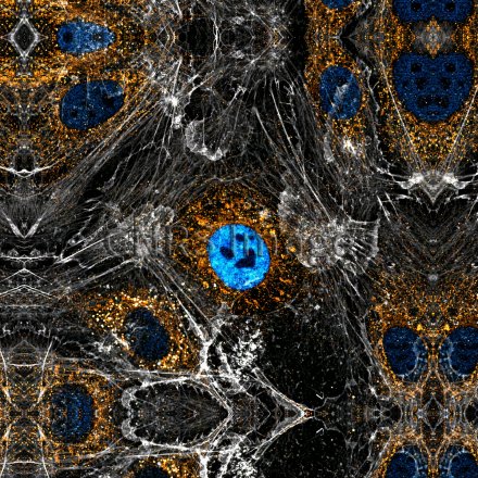
To invade other organs, tumour cells engage in a very sophisticated dialogue with the surrounding tissue and the extracellular matrix. In particular, they trigger the creation of new blood vessels (angiogenesis) to obtain food and proliferate. Here, it is precisely these reprogramming mechanisms that scientists are trying to identify using endothelial cells, which line the inside of vessels. In this complex microenvironment, their structure stands out, in grey, due to their actin cytoskeleton. An oncofoetal form of fibronectin, a protein that plays a key role in cell adhesion to the extracellular matrix, is shown in orange. Present in embryonic life, this form of fibronectin is expressed again during tumour progression, promoting cancer angiogenesis. And finally, in blue in the cell nucleus, protein SAM68 is suspected of being involved in the regulation of oncofoetal fibronectin. Excluded from certain regions of the nucleus, it sometimes produces smiley-like forms, such as the one featured here, with a heart-shaped mouth. This image is a winner of the 2021 La preuve par l'image (LPPI) competition.
The use of media visible on the CNRS Images Platform can be granted on request. Any reproduction or representation is forbidden without prior authorization from CNRS Images (except for resources under Creative Commons license).
No modification of an image may be made without the prior consent of CNRS Images.
No use of an image for advertising purposes or distribution to a third party may be made without the prior agreement of CNRS Images.
For more information, please consult our general conditions
2020
Our work is guided by the way scientists question the world around them and we translate their research into images to help people to understand the world better and to awaken their curiosity and wonderment.