Production year
2017

© Orestis FAKLARIS / Nicolas CHEVALIER / IJM / CEMIBIO / CNRS Images
20180032_0012
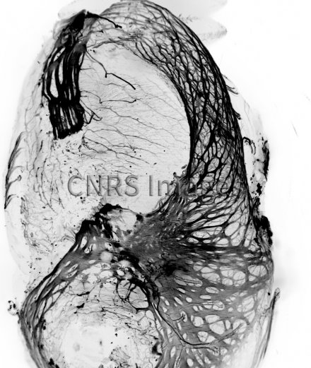
Projection en Z du système nerveux de l'estomac et du système digestif d’un embryon de poulet de 15 jours, observée en microscopie à feuille de lumière. Les marquages utilisés sont la Beta-Tubuline de classe III couplée à une Alexa Fluor 488. L’échantillon a été rendu transparent, par la méthode iDISCO+, afin de pouvoir imager l’échantillon entier en 3D, sans devoir effectuer de coupes. La transparisation (ou clarification) permet ici de révéler les plexus neuronaux même en profondeur.
Cette image a été soumise à l’édition 2017 du concours d’images de France-Bioimaging (UMS3714-CEMIBIO, CNRS)
The use of media visible on the CNRS Images Platform can be granted on request. Any reproduction or representation is forbidden without prior authorization from CNRS Images (except for resources under Creative Commons license).
No modification of an image may be made without the prior consent of CNRS Images.
No use of an image for advertising purposes or distribution to a third party may be made without the prior agreement of CNRS Images.
For more information, please consult our general conditions
2017
Our work is guided by the way scientists question the world around them and we translate their research into images to help people to understand the world better and to awaken their curiosity and wonderment.