Production year
2017

© Cyril FRESILLON / PPRIME / CNRS Images
20170142_0041
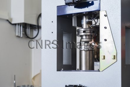
Échantillon installé entre les plateaux de compression sur la machine d'essai mécanique in-situ d'un tomographe. La micro-tomographie est une puissante technique d'imagerie 3D permettant notamment d'étudier le comportement de différentes classes de matériaux (polymères, composites, métaux...) soumis à des sollicitations de nature diverse (mécanique, thermique...). La caractérisation de l'endommagement subi par ces matériaux peut se réaliser post-mortem mais la compréhension des différents mécanismes est facilitée par la réalisation d'essais in situ. Il s'agit ici de caractériser l'endommagement d'un échantillon de composite carbone/époxy soumis à une sollicitation de compression.
The use of media visible on the CNRS Images Platform can be granted on request. Any reproduction or representation is forbidden without prior authorization from CNRS Images (except for resources under Creative Commons license).
No modification of an image may be made without the prior consent of CNRS Images.
No use of an image for advertising purposes or distribution to a third party may be made without the prior agreement of CNRS Images.
For more information, please consult our general conditions
2017
Our work is guided by the way scientists question the world around them and we translate their research into images to help people to understand the world better and to awaken their curiosity and wonderment.