© Lionel CHASSON / CIML / CNRS Images
Reference
20170103_0020
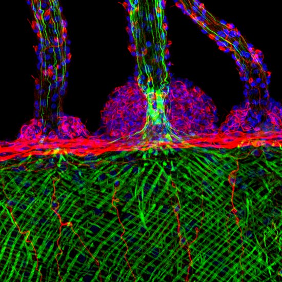
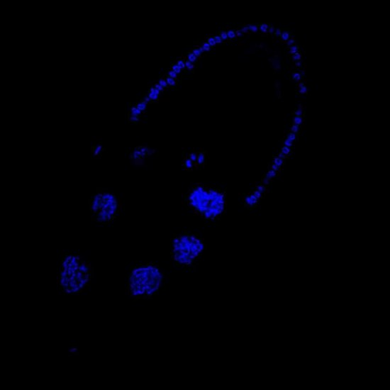
Coupe de rate de souris observée en microscopie confocale multi-couleur
Cross-section of a mouse spleen, observed using multicolour confocal microscopy. Immunofluorescent marking reveals B lymphocytes (in blue) and two sub-populations of T lymphocytes (in green and red). The image was obtained by combining several lasers, including a white-light laser that generates a wide range of wavelengths and hence colours. A high-power oil-immersion objective lens was used. With confocal microscopy, laser beams scan the specimen at high speed, point by point. This technique can be used to produce thin optical cross-sections (at various altitudes within a thick specimen) that reveal the locations of numerous entities and cell types (T and B lymphocytes, dendrites, macrophages, actins, nuclei, etc.) in a tissue slice. These optical cross-sections are then stacked to generate 3D images of the specimens. This enables scientists to compare the locations and numbers of entities in various circumstances, such as in patients suffering from diseases, for example, in order to shed light on the workings of the immune system. This technique is now widely used in laboratories, and is constantly being improved in terms of its resolution and sensitivity, as well as the possible colours and contrasts.
Add to my selection
Terms of use
The use of media visible on the CNRS Images Platform can be granted on request. Any reproduction or representation is forbidden without prior authorization from CNRS Images (except for resources under Creative Commons license).
No modification of an image may be made without the prior consent of CNRS Images.
No use of an image for advertising purposes or distribution to a third party may be made without the prior agreement of CNRS Images.
For more information, please consult our general conditions