Production year
2006

© Laurent VANHILLE / CMIL / INSERM / CNRS Images
20170103_0006
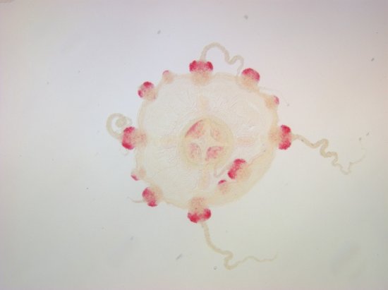
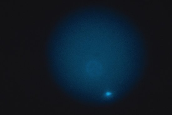
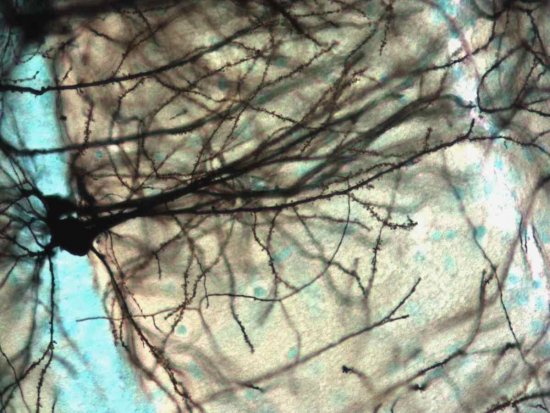
Macrophages in the peritoneum of mice deficient in the MafB transcription factor, exhibiting significant morphological modifications. These cells were cultivated for five minutes in a medium containing M-CSF (a cytokine), and were fixed and coloured using a phalloidin combined with TRITC (in red) to reveal the actin organisation. The MafB-deficient macrophages have prominent protuberances and are often branched. It was long thought that production of the various specialist cells from stem cells in the blood occurred randomly. However, scientists have discovered that in the case of myeloid cells (stem cells that evolve into leukocytes, of which macrophages are one form), the decisive factor is the combined action of two different proteins, one located inside the cell (a MafB transcription factor), the other outside it (the cytokine M-CSF). Transcription factors activate or inhibit genes, thereby determining a cell's identity.
The use of media visible on the CNRS Images Platform can be granted on request. Any reproduction or representation is forbidden without prior authorization from CNRS Images (except for resources under Creative Commons license).
No modification of an image may be made without the prior consent of CNRS Images.
No use of an image for advertising purposes or distribution to a third party may be made without the prior agreement of CNRS Images.
For more information, please consult our general conditions
2006
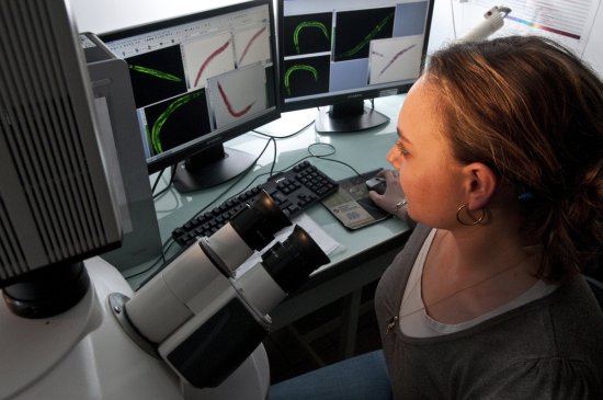
Our work is guided by the way scientists question the world around them and we translate their research into images to help people to understand the world better and to awaken their curiosity and wonderment.