Production year
2017

© Hugues LELOUARD / CIML / INSERM / CNRS Images
20170103_0001
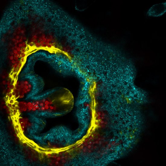
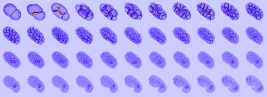
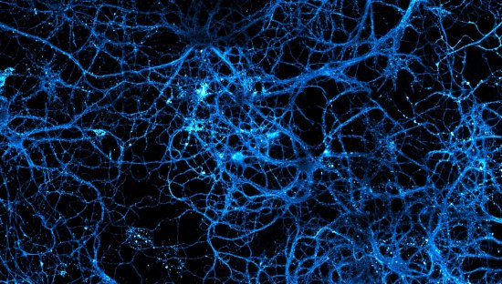
Small intestine of a mouse, observed using multicolour confocal microscopy. Immunofluorescent marking reveals epithelial cells in cyan, blood vessels in magenta, lymph vessels in orange, phagocytic cells in red, monocytes in green, paneth cells and mucus in grey, collagen fibres in yellow, and lastly, actin filaments in purple. The image was obtained by combining several lasers and a spectral detector able to distinguish a wide range of fluorochromes, i.e. colours. A high-power (40X) oil-immersion objective lens was used. With confocal microscopy, laser beams scan the specimen at high speed, point by point. This technique can be used to produce thin optical cross-sections (at various specimen thicknesses) that reveal the locations of numerous entities and cell types (T and B lymphocytes, dendrites, macrophages, actins, nuclei, etc.) in a tissue slice. These optical cross-sections are then stacked to generate 3D images of the specimens. This enables scientists to compare the locations and numbers of entities in various circumstances, such as in patients suffering from diseases, for example, in order to shed light on the workings of the immune system. This technique is constantly being improved in terms of its resolution and sensitivity, as well as the possible colours and contrasts.
The use of media visible on the CNRS Images Platform can be granted on request. Any reproduction or representation is forbidden without prior authorization from CNRS Images (except for resources under Creative Commons license).
No modification of an image may be made without the prior consent of CNRS Images.
No use of an image for advertising purposes or distribution to a third party may be made without the prior agreement of CNRS Images.
For more information, please consult our general conditions
2017
Our work is guided by the way scientists question the world around them and we translate their research into images to help people to understand the world better and to awaken their curiosity and wonderment.