© Véronique GASSER / BSC / UNISTRA / CNRS Images
Reference
20170099_0016
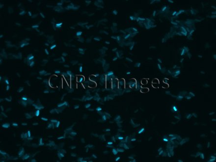
Bacille pyocyanique observé en microscopie optique à fluorescence
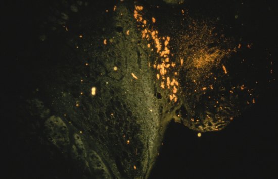
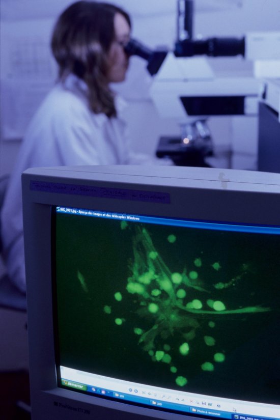
Co-marking DNA breaks in HeLa cells produced by a laser, observed using fluorescence microscopy. The breaks are shown in yellow, as a result of co-marking with the red colour of the breaks mixed with the green of the protein XRRC1, which bonds to them. Here, scientists are analysing the distribution of the DNA breaks across various types of cells, in order to determine which repair pathways are active, and identify any possible interactions between them. Cancer cells have a high proportion of breaks in their DNA. Therapeutic agents used in cancer treatment inhibit the repair pathways for such breaks.
The use of media visible on the CNRS Images Platform can be granted on request. Any reproduction or representation is forbidden without prior authorization from CNRS Images (except for resources under Creative Commons license).
No modification of an image may be made without the prior consent of CNRS Images.
No use of an image for advertising purposes or distribution to a third party may be made without the prior agreement of CNRS Images.
For more information, please consult our general conditions

