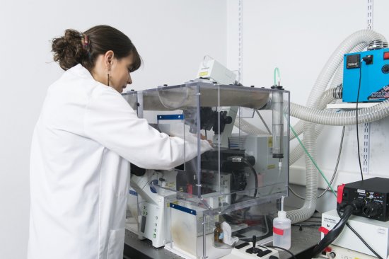
© Jean-Christophe AME / BSC / UNISTRA / CNRS Images
Reference
20170099_0013
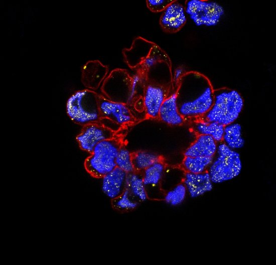
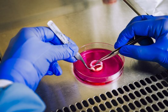
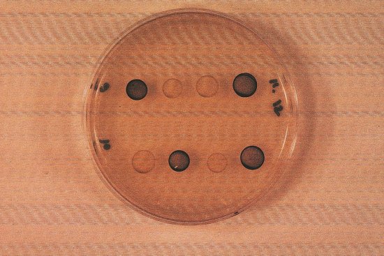
Métaphase anormale de Cellules HeLa
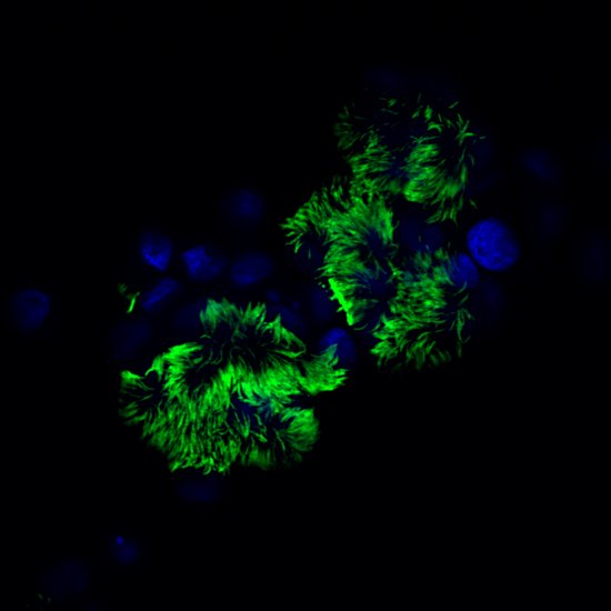
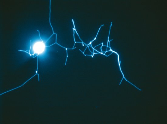
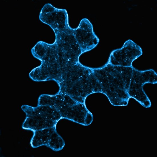
Interphase HeLa cell observed using fluorescence microscopy. DNA is marked in blue, histone H3 in green, and cytochrome C (released by mitochondria upon cell death) in red. The cell volume increases during the interphase, immediately prior to division. Here, researchers are studying a particular case of cell death, known as mitotic catastrophe. This phenomenon may be caused by a defect in the cell cycle sequence or by a DNA alteration. More specifically, the scientists are interested in cells that have lost the PARG function. This enzyme - Poly (ADP-ribose) glycohydrolase (PARG) - degrades poly(ADP-ribose), a small branched molecule that temporarily modifies proteins with a role in repairing damage to DNA.
Add to my selection
Terms of use
The use of media visible on the CNRS Images Platform can be granted on request. Any reproduction or representation is forbidden without prior authorization from CNRS Images (except for resources under Creative Commons license).
No modification of an image may be made without the prior consent of CNRS Images.
No use of an image for advertising purposes or distribution to a third party may be made without the prior agreement of CNRS Images.
For more information, please consult our general conditions