Production year
2004

© Andrea PASINI / IBDM / CNRS Images
20170090_0003
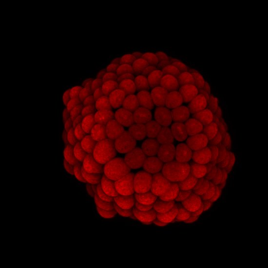
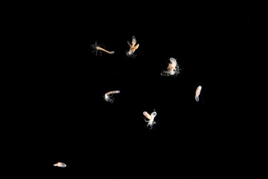
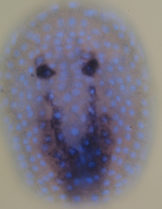
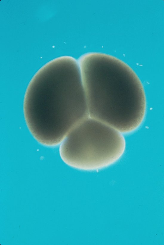
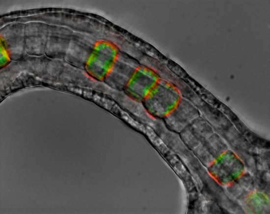
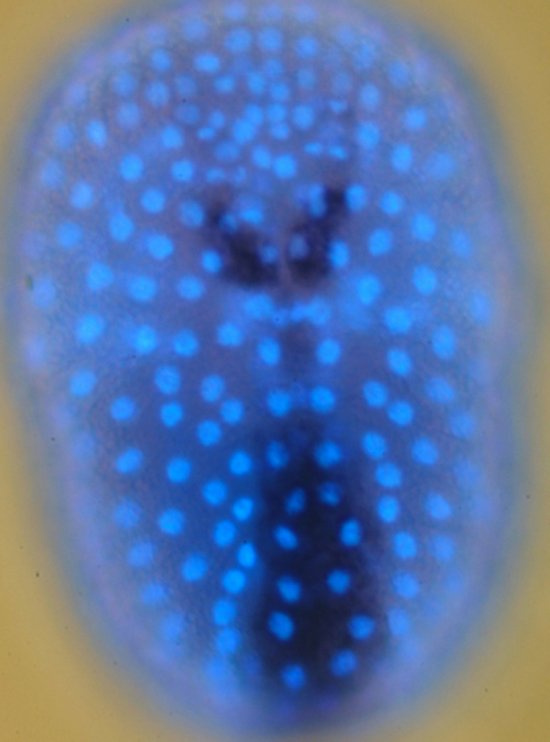
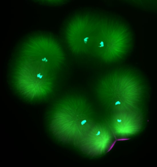
Embryo of the solitary sea squirt Ciona intestinalis (Chordata, Tunicata), a benthic marine invertebrate and plankton filter-feeder, observed using an epifluorescence microscope. The embryos have been coloured using a DNA-marker (light blue) to highlight the cell nuclei, and various RNA probes (dark blue/black), in order to uncover the expression profile of the genes involved in the development of the epidermis and peripheral nervous system. Its relative anatomical and genetic simplicity, as well as its phylogenetic similarity to vertebrates, make Ciona intestinalis a commonly used model organism for the study of embryo development.
The use of media visible on the CNRS Images Platform can be granted on request. Any reproduction or representation is forbidden without prior authorization from CNRS Images (except for resources under Creative Commons license).
No modification of an image may be made without the prior consent of CNRS Images.
No use of an image for advertising purposes or distribution to a third party may be made without the prior agreement of CNRS Images.
For more information, please consult our general conditions
2004
Our work is guided by the way scientists question the world around them and we translate their research into images to help people to understand the world better and to awaken their curiosity and wonderment.