© Liu ZENG ZHEN / IJM / CNRS Images
Reference
20170082_0007
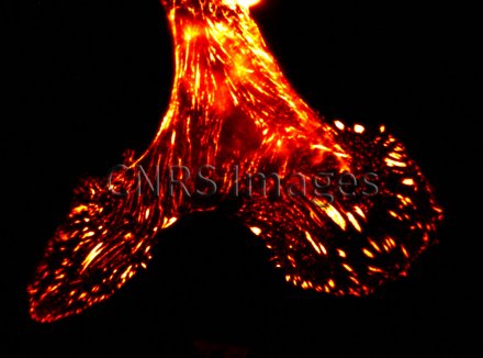
Protéines d'adhésion dans une cellule souche en microscopie de fluorescence confocale
Adhesion proteins in a stem cell, observed using a Zeiss LSM 700 fluorescence confocal microscope. In confocal microscopy, the sample is scanned by laser beams, point by point. This technique produces fine optical sections (at different levels in a thick sample) that reveal the location of numerous entities or cell types in a section of tissue or individual cells. These optical sections are then stacked to generate 3D images of samples. This technique, used commonly in laboratories, is improving all the time in terms of the resolution, sensitivity, colours and contrasts possible.
The use of media visible on the CNRS Images Platform can be granted on request. Any reproduction or representation is forbidden without prior authorization from CNRS Images (except for resources under Creative Commons license).
No modification of an image may be made without the prior consent of CNRS Images.
No use of an image for advertising purposes or distribution to a third party may be made without the prior agreement of CNRS Images.
For more information, please consult our general conditions