© Sébastien MAILFERT / Noushin MOSSADEGH / CIML / INSERM / AMU / CNRS Images
Reference
20170082_0003
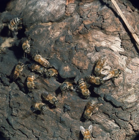
Coupe de testicule de souris en microscopie confocale multi-couleur
Section of a seminiferous tubule from a mouse testis measuring 20 µm thick, observed using a multicolour confocal microscope. The cell nuclei are visible in cyan (DAPI), with the actin, which reveals the cell skeleton, in red (phalloidin labelled with the fluorochrome Alexa-647) and the macrophages in green Testicular macrophages are mobilised to defend the spermatozoa. By emitting specific molecules, these fertility guardians prevent other elements in the immune system entering the testes. A recent study made it possible to characterise the two types of testicular macrophage and find out about the origin, development and characteristics of these immune cells. The first type of testicular macrophage is of embryonic origin, while the peritubular macrophages are located around the seminiferous tubules. By using a new cell tracing method, researchers were able to follow the peritubular macrophages (from the bone marrow to the testes), which only appear two weeks after the mice are born, the stage that corresponds to puberty in humans. Surprisingly, once established in the testes, both macrophage populations remain there for the rest of their lives. This discovery could lead to the development of new treatments for male infertility. In confocal microscopy, the technique used to produce this image, the sample is scanned by laser beams, point by point. This produces fine optical sections that reveal the location of numerous entities or cell types (T cells, B cells, dendritic cells, macrophages, actin, nucleus, etc.) in a section of tissue. In this way, research scientists can compare the number and location of entities in various situations, in individuals affected by pathologies, for example, in order to decipher the mechanisms of the immune system. This technique, used commonly in laboratories, is improving all the time in terms of the resolution, sensitivity, colours and contrasts possible.
Add to my selection
Terms of use
The use of media visible on the CNRS Images Platform can be granted on request. Any reproduction or representation is forbidden without prior authorization from CNRS Images (except for resources under Creative Commons license).
No modification of an image may be made without the prior consent of CNRS Images.
No use of an image for advertising purposes or distribution to a third party may be made without the prior agreement of CNRS Images.
For more information, please consult our general conditions