© Sébastien MAILFERT / CIML / INSERM / AMU / CNRS Images
Reference
20170082_0001
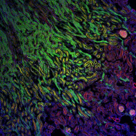
Section de rein de souris observée en microscopie confocale multi-couleur
Section of a mouse kidney observed using a multicolour confocal microscope. This microscope image, which has a resolution of around 200 nanometres, was obtained using immunofluorescence labelling: AF488-WGA in green, AF568 phalloidin in red and DAPI in blue. The image was generated using a combination of several lasers, including one white laser providing a wide range of wavelengths (colours). In confocal microscopy, the sample is scanned by laser beams, point by point. This technique produces fine optical sections (at different levels in a thick sample) that reveal the location of numerous entities or cell types (T cells, B cells, dendritic cells, macrophages, actin, nucleus, etc.) in a section of tissue. These optical sections are then stacked to generate 3D images of samples. In this way, research scientists can compare the number and location of entities in various situations, in individuals affected by pathologies, for example, in order to decipher the mechanisms of the immune system. This technique, used commonly in laboratories, is improving all the time in terms of the resolution, sensitivity, colours and contrasts possible.
The use of media visible on the CNRS Images Platform can be granted on request. Any reproduction or representation is forbidden without prior authorization from CNRS Images (except for resources under Creative Commons license).
No modification of an image may be made without the prior consent of CNRS Images.
No use of an image for advertising purposes or distribution to a third party may be made without the prior agreement of CNRS Images.
For more information, please consult our general conditions


