Production year
2017

© Hubert RAGUET / Institut Cochin / CNRS Images
20170080_0012
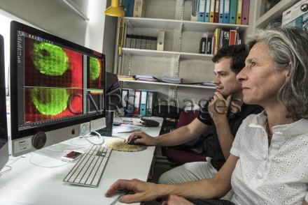
Characterisation of the infection of mucosal cells with HIV, recorded in real-time using a microscope and analysed using the Imaris software package. To view the different elements involved in infection, the cells producing the virus have been modified to produce a fluorescent virus that contains green fluorescent proteins (GFP), while the genital mucosal cells have been genetically modified so that they express a red fluorescent protein (RFP) on the surface. The research scientists then reproduce, in vivo, the first stage of the mucosal entry of the virus, i.e. the transfer of HIV from an infected cell to a male genital cell. Watching this in real time using the microscope take one hour, during which time the cells (infected and target) are scanned continuously. By superimposing around 100 sections of these scans, the scientists obtain a reconstruction in space and time (4D). This has made it possible to demonstrate that infected cells, when they come into contact with the epithelial cells, form a tight connection with the target mucosal cells. This is referred to as a viral synapse, which stimulates local production of the virus. The virus can then cross the epithelial barrier via transcytosis, a transcellular transportation mechanism. This viral synapse causes the mucosal cell to secrete cytokines, including thymic stromal lymphopoietin (TSLP), which in this case acts as a chemokine. Chemokines are proteins that control the positioning of immune cells in tissues. The TSLP secreted by these mucosal cells attracts Langerhans cells towards the lumen of the mucosa. These Langerhans cells then capture the virus and start the infection proper. The "Mucosal entry of HIV and mucosal immunity" team led by Morgane Bomsel is attempting to target TSLP in order to develop a prophylactic treatment that prevents the mucosal transmission of the virus, which is the main entry route of HIV into the organism.
The use of media visible on the CNRS Images Platform can be granted on request. Any reproduction or representation is forbidden without prior authorization from CNRS Images (except for resources under Creative Commons license).
No modification of an image may be made without the prior consent of CNRS Images.
No use of an image for advertising purposes or distribution to a third party may be made without the prior agreement of CNRS Images.
For more information, please consult our general conditions
2017
Our work is guided by the way scientists question the world around them and we translate their research into images to help people to understand the world better and to awaken their curiosity and wonderment.