Production year
2016

© Caroline FONTA/Franck PLOURABOUE/CERCO/IMFT/CNRS Images
20170064_0001
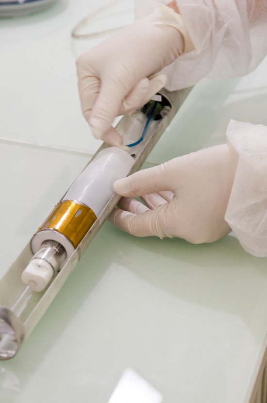
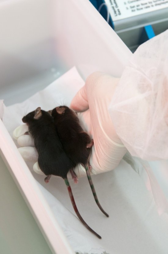
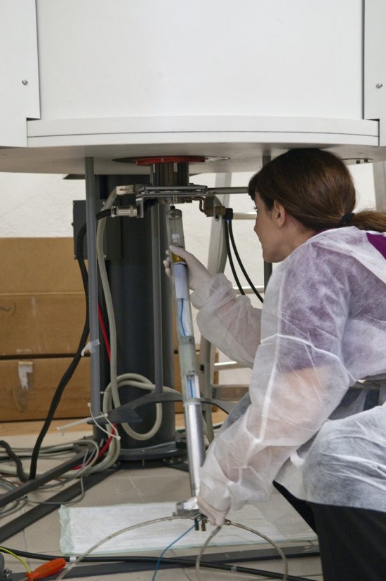
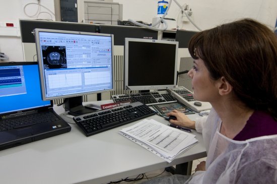
Reconstruction of the microcirculatory system in a sample measuring a few square millimetres from the cerebral cortex of a primate, using X-ray tomography (European Synchrotron Radiation Facility). This model was obtained by injecting a contrast product, barium sulphate, into the system. The top of the image corresponds to the surface of the brain, where we can see the dense, high-volume networks that characterise grey matter. White matter, on the other hand, can be identified by the finer networks, which appear at the bottom of the image. The clear separation between the two types of network is a cortical fold. Tomography makes it possible to explore, with great precision, the way in which microcirculation is organised, with its small vessels that supply cells with oxygen. Research scientists have performed a quantitative study of this system and found that the networks are larger and more distant in the case of a tumour, which may explain why certain treatments fail. This type of modelling is also used to produce digital simulations of blood flow or blood clots in the bloodstream.
The use of media visible on the CNRS Images Platform can be granted on request. Any reproduction or representation is forbidden without prior authorization from CNRS Images (except for resources under Creative Commons license).
No modification of an image may be made without the prior consent of CNRS Images.
No use of an image for advertising purposes or distribution to a third party may be made without the prior agreement of CNRS Images.
For more information, please consult our general conditions
2016
Our work is guided by the way scientists question the world around them and we translate their research into images to help people to understand the world better and to awaken their curiosity and wonderment.