Production year
2015

© Yvon JAILLAIS/RDP/CNRS Images
20170028_0001
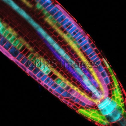
Montage of confocal microscopy images showing a root tip of thale cress, Arabidopsis thaliana, whose cell membranes are marked by a red fluorescent dye. Each cell line is established under the influence of auxin, a hormone that acts like the conductor of an orchestra in terms of root development, determining the position and rate of growth of new roots. These lines are revealed by the expression of a fluorescent protein of a different colour in each root tissue: lateral calyptra and cortex (magenta and green), columella and protoxylem (cyan), epidermis and protophloem (blue), endoderm (yellow), quiescent centre (orange). These markers are used to study root development and its response to environmental changes.
The use of media visible on the CNRS Images Platform can be granted on request. Any reproduction or representation is forbidden without prior authorization from CNRS Images (except for resources under Creative Commons license).
No modification of an image may be made without the prior consent of CNRS Images.
No use of an image for advertising purposes or distribution to a third party may be made without the prior agreement of CNRS Images.
For more information, please consult our general conditions
2015
Our work is guided by the way scientists question the world around them and we translate their research into images to help people to understand the world better and to awaken their curiosity and wonderment.