© Virginie CALLOT/CRMBM-CEMEREM/CNRS Images
Reference
20160112_0006
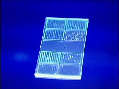
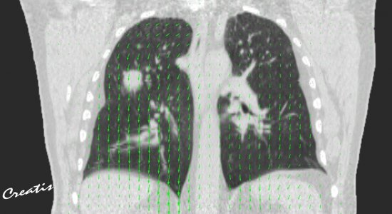
Cartography of the spinal cord in a healthy subject
Cartography of the spinal cord in a healthy subject using high spatial resolution T1-weighted image contrast (0.6 mm isotropic). This image is a cartography of the longitudinal relaxation time (T1) in the sagittal plane, derived from 7-tesla MRI with a T1-weighted image. Quantitative MRI enables us to investigate structural differences in the different tissue compartments (grey matter, white matter, types of white matter fibre bundles). It can also detect pathological anomalies in comparison with average control values. Finally, it can be used as a biomarker and help in the objective assessment of therapeutic efficiency.
Add to my selection
Terms of use
The use of media visible on the CNRS Images Platform can be granted on request. Any reproduction or representation is forbidden without prior authorization from CNRS Images (except for resources under Creative Commons license).
No modification of an image may be made without the prior consent of CNRS Images.
No use of an image for advertising purposes or distribution to a third party may be made without the prior agreement of CNRS Images.
For more information, please consult our general conditions