© Virginie CALLOT/CRMBM-CEMEREM/CNRS Images
Reference
20160112_0005
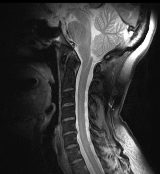
Very high spatial resolution morphological image of a spinal cord at cervical level, acquired by MRI
Very high spatial resolution morphological image (0.18 x 0.18 mm2 in the plane) of a spinal cord at cervical level, acquired by ultra-high field (7-tesla) magnetic resonance imaging (MRI). The contrast obtained enables us to distinguish the grey matter clearly from the white matter. Sub-structures can also be identified within the white matter (slender and wedge-shaped bundles). Other details such as nerve roots, blood vessels and ligaments are also visible. Thanks to improved signal-to-noise ratios and new contrast levels, ultra-high field MRI enables us to see structures that until now were barely visible on conventional 1.5- and 3-tesla imaging devices, thus opening up new perspectives for a much finer characterisation of tissues and their alterations.
Add to my selection
Terms of use
The use of media visible on the CNRS Images Platform can be granted on request. Any reproduction or representation is forbidden without prior authorization from CNRS Images (except for resources under Creative Commons license).
No modification of an image may be made without the prior consent of CNRS Images.
No use of an image for advertising purposes or distribution to a third party may be made without the prior agreement of CNRS Images.
For more information, please consult our general conditions