Production year
2011

© Claude SAUTER/IBMC/CNRS Images
20140001_1139
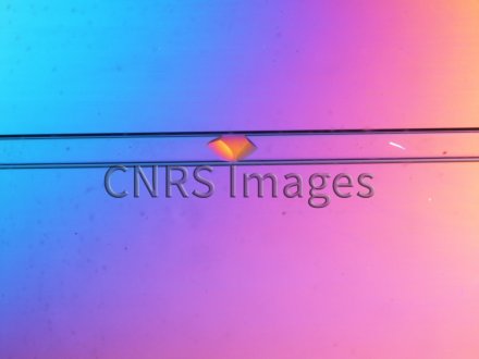
Cristallisation d'une protéine en tube capillaire (en vue d'études structurales par cristallographie). L'analyse d'un cristal par diffraction des rayons permet de visualiser la protéine empilée au sein de ce cristal. Il en résulte une image 3D de la protéine qui renseigne sur sa forme, mais également sur sa fonction biologique. Il s'agit ici d'une enzyme (ou biocatalyseur) appelée lysozyme qui coupe l'enveloppe de sucre à la surface de certaines bactéries.
The use of media visible on the CNRS Images Platform can be granted on request. Any reproduction or representation is forbidden without prior authorization from CNRS Images (except for resources under Creative Commons license).
No modification of an image may be made without the prior consent of CNRS Images.
No use of an image for advertising purposes or distribution to a third party may be made without the prior agreement of CNRS Images.
For more information, please consult our general conditions
2011
Our work is guided by the way scientists question the world around them and we translate their research into images to help people to understand the world better and to awaken their curiosity and wonderment.