Production year
2011

© Cyril FRESILLON/CNRS Images
20120001_0021
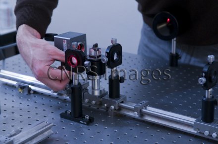
Réglage de l'angle d'incidence de l'illumination de référence sur la caméra pour optimiser l'enregistrement des interférogrammes. Microscope interférométrique permettant de mesurer l'amplitude et la phase de la lumière diffractée par l'objet étudié. L'objet est illuminé successivement selon différents angles pour reconstruire par tomographie la cartographie de son indice de réfraction en trois dimensions. L'application est l'imagerie 3D haute résolution, utilisée en biologie et pour la caractérisation de nanocomposants.
The use of media visible on the CNRS Images Platform can be granted on request. Any reproduction or representation is forbidden without prior authorization from CNRS Images (except for resources under Creative Commons license).
No modification of an image may be made without the prior consent of CNRS Images.
No use of an image for advertising purposes or distribution to a third party may be made without the prior agreement of CNRS Images.
For more information, please consult our general conditions
2011
Our work is guided by the way scientists question the world around them and we translate their research into images to help people to understand the world better and to awaken their curiosity and wonderment.