Production year
2017

© Claire WILHELM / MSC / CNRS Images
20170105_0001
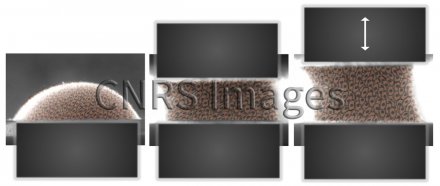
Embryonic stem cells stimulated mechanically using a magnetic tissue stretcher. This device shapes and mechanically stimulates an aggregate of cells into which magnetic nanoparticles have been incorporated. The two micro-magnets, one of which is mobile, are placed either side of the resulting embryoid body, and the cyclical stimulation can be adjusted according to the type of tissue to be produced. Scientists observed that when the stimulation process generated magnetic pulses mimicking heart beats, stem cells differentiated into cardiac muscle cell precursors. This discovery addresses one of the current challenges in regenerative medicine, by making it possible to create an organised, cohesive cell assembly with no need for an extracellular matrix. Researchers have also proved that incorporating nanoparticles affects neither the function nor the differentiation capabilities of stem cells. This device is opening up new fields in biophysics research and tissue engineering.
The use of media visible on the CNRS Images Platform can be granted on request. Any reproduction or representation is forbidden without prior authorization from CNRS Images (except for resources under Creative Commons license).
No modification of an image may be made without the prior consent of CNRS Images.
No use of an image for advertising purposes or distribution to a third party may be made without the prior agreement of CNRS Images.
For more information, please consult our general conditions
2017
Our work is guided by the way scientists question the world around them and we translate their research into images to help people to understand the world better and to awaken their curiosity and wonderment.