Production year
2017

© Boris BARBOUR/IBENS/CNRS Images
20170011_0001
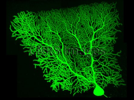
View under confocal microscope of an adult rat Purkinje cell marked selectively by means of a fluorescent molecule, after patch-clamp recording (technique consisting of recording the electrical activity of a microscopic fragment of cell membrane. The cell body (round shape at the bottom of the image) measures approximately 25 microns in diameter and the dendritic tree spans approximately 200 x 200 microns, but it is almost flat. Terminal dendrites, known as thorny twigs, bear numerous dendritic spines, sorts of small mushrooms of approximately 1 micron where synaptic contacts are established. Purkinje cells receive approximately 200,000 synapses, whose modification by synaptic plasticity enables storage of the information acquired during motor learning. These are the main cells of the cerebellar cortex. The cerebellum plays a key role in learning, refinement and automation of coordinated movements.
The use of media visible on the CNRS Images Platform can be granted on request. Any reproduction or representation is forbidden without prior authorization from CNRS Images (except for resources under Creative Commons license).
No modification of an image may be made without the prior consent of CNRS Images.
No use of an image for advertising purposes or distribution to a third party may be made without the prior agreement of CNRS Images.
For more information, please consult our general conditions
2017
Our work is guided by the way scientists question the world around them and we translate their research into images to help people to understand the world better and to awaken their curiosity and wonderment.