Production year
2015

© Katalin CZONDOR/Olivier THOUMINE/IINS/CNRS Images
20170022_0010
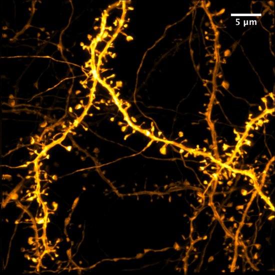
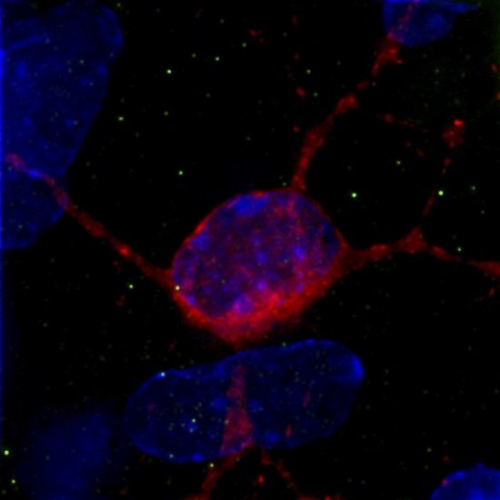
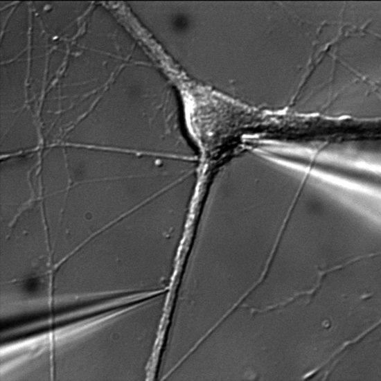
Neuron expressing actin fused to green fluorescent protein (GFP), in dissociated hippocampal neurons (orange marking) in rats. It reveals actin enrichment in the dendritic spines, these small protrusions that represent the excitatory post-synapses. The phalloidin in blue is a filamentous actin marker in the surrounding neurons. This image was produced by confocal microscopy.
The use of media visible on the CNRS Images Platform can be granted on request. Any reproduction or representation is forbidden without prior authorization from CNRS Images (except for resources under Creative Commons license).
No modification of an image may be made without the prior consent of CNRS Images.
No use of an image for advertising purposes or distribution to a third party may be made without the prior agreement of CNRS Images.
For more information, please consult our general conditions
2015
Our work is guided by the way scientists question the world around them and we translate their research into images to help people to understand the world better and to awaken their curiosity and wonderment.