Folder

Cell biology: breaking down the boundaries of the visible world

The progress in imaging techniques is inseparable from the advances in knowledge in cell biology, improving our understanding of the cell and intercellular interactions.

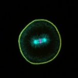
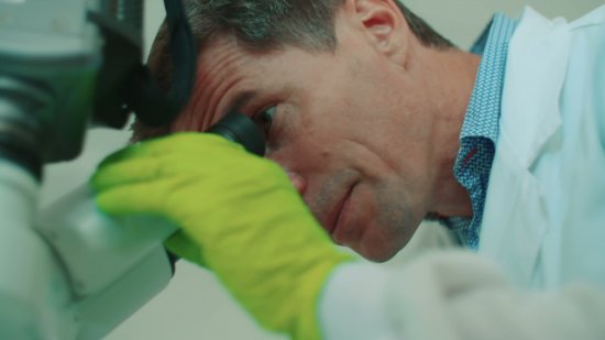
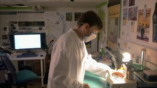
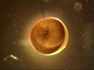
Observation et acquisition d’images de cellules neurales, au microscope à fluorescence.

© Cyril Frésillon / CNRS Photothèque
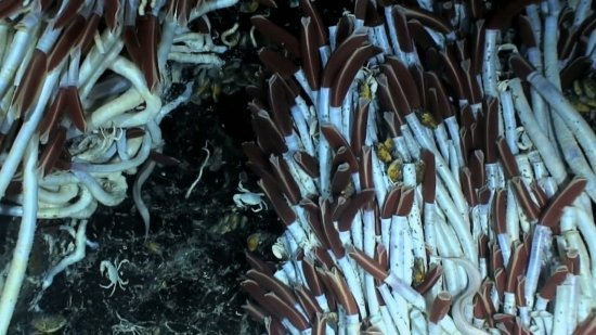
View the media

Advances in imaging technologies and biophysics have facilitated considerable breakthroughs in observing cells, their components and behaviour. They have helped us to improve our knowledge of different cell types, their functions, but also how they divide and communicate through contact or remotely.

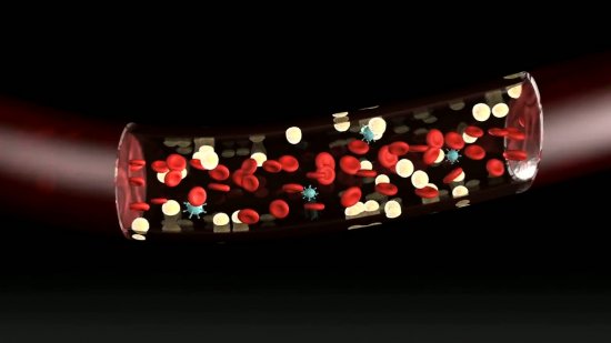
Optical microscopes, which use a light beam, have paved the way for electron microscopes, which use an electron beam, and for confocal microscopy, which uses a laser. Their resolution shows the external and internal structure of organelles with precision. When combined with the use of biological markers, these types of microscopes are excellent tools for identifying extracellular transport and intracellular signalling pathways.

More recently, super-resolution microscopy and the high-throughput screening of 3D biological objects are breaking down new resolution barriers. In addition to observation, it is now possible to manipulate cells individually using remote-controlled microrobots with optical tweezers.

Our increasingly detailed knowledge of the cellular machinery is even leading scientists to design artificial cells, or to reprogramme cells to perform completely different tasks to their natural functions, which opens the door to many applications.

Discover in pictures an overview of the cell biology research undertaken in the CNRS laboratories.

Key words: biophysics, cell, cellular engineering, confocal, imaging, microfluidics, optical tweezers, SEM (scanning electron microscope), TEM (transmission electron microscope)

Open media modal

Only available for non-commercial distribution

For any commercial distribution contact the executive producer

Ifremer teams have found a bacterium at the bottom of the oceans that could contribute to the development of regenerative medicine. The aim of teams from the Inserm 791 “Osteo-articular and dental engineering laboratory” research unit and Ifremer is to manufacture injectable gels capable of stimulating the regeneration of cartilage or bone from stem cells. Their key ingredient is a polysaccharide produced by a bacteria of marine origin called Alteromonas infernus.

Video
4864
Restorative bacteria
Open media modal

Only available for non-commercial distribution

Concepteur de cellules artificielles, Franck Molina reprogramme le vivant pour des applications de diagnostic. Il obtient ainsi des détecteurs rapides, bon marché, qui changent de couleur en présence de certains biomarqueurs. Ses travaux ont fait l'objet de nombreux transferts industriels. Directeur de recherche CNRS au laboratoire Modélisation et ingénierie des systèmes complexes biologiques pour le diagnostic (Sys2Diag), qu'il dirige, Franck Molina est un…

Video
7352
Médaille de l'innovation 2020 : Franck Molina, chercheur en biotechnologies
Open media modal

Only available for non-commercial distribution

Portrait de Vincent Baylé, lauréat de la Médaille de Cristal 2020 du CNRS, ingénieur de recherche en microscopie et traitement d'images, spécialiste en microscopie et traitement d'images au sein de l'équipe Signalisation cellulaire et endocytose du laboratoire Reproduction et développement des plantes

Video
6993
Médaille de Cristal 2020 : Vincent Baylé, ingénieur de recherche en microscopie et traitement d'images
Open media modal

The CNRS Gold Medal, France's most prestigious scientific distinction, has been awarded this year to the developmental biologist Margaret Buckingham, exceptional grade senior researcher emeritus at CNRS and professor emeritus at the Institut Pasteur. Her research work has led to important advances in the field of myogenesis (muscle formation), cardiogenesis (formation of the heart) and stem cells in embryos and adults. In addition to their contribution to fundamental knowledge, her discoveries…

Video
4157
Margaret Buckingham, a biologist fascinated by development
Open media modal

There are continual exchanges between a cell's cytoplasm and its nucleus. In 1997, a team led by Catherine Dargemont of the Institut Jacques Monod was the first to develop a method for extracting proteins from the nucleus. Since then this laboratory has focused on several areas: the mechanisms regulating the exchanges; the mechanisms for transporting RNA and in particular the stage of translocation of the transport complexes in the cytoplasm; understanding how certain viruses like HIV make use…

Video
1056
Goings-on inside the cell

CNRS Images,

Our work is guided by the way scientists question the world around them and we translate their research into images to help people to understand the world better and to awaken their curiosity and wonderment.